Copyright
©2013 Baishideng Publishing Group Co.
World J Radiol. Nov 28, 2013; 5(11): 421-429
Published online Nov 28, 2013. doi: 10.4329/wjr.v5.i11.421
Published online Nov 28, 2013. doi: 10.4329/wjr.v5.i11.421
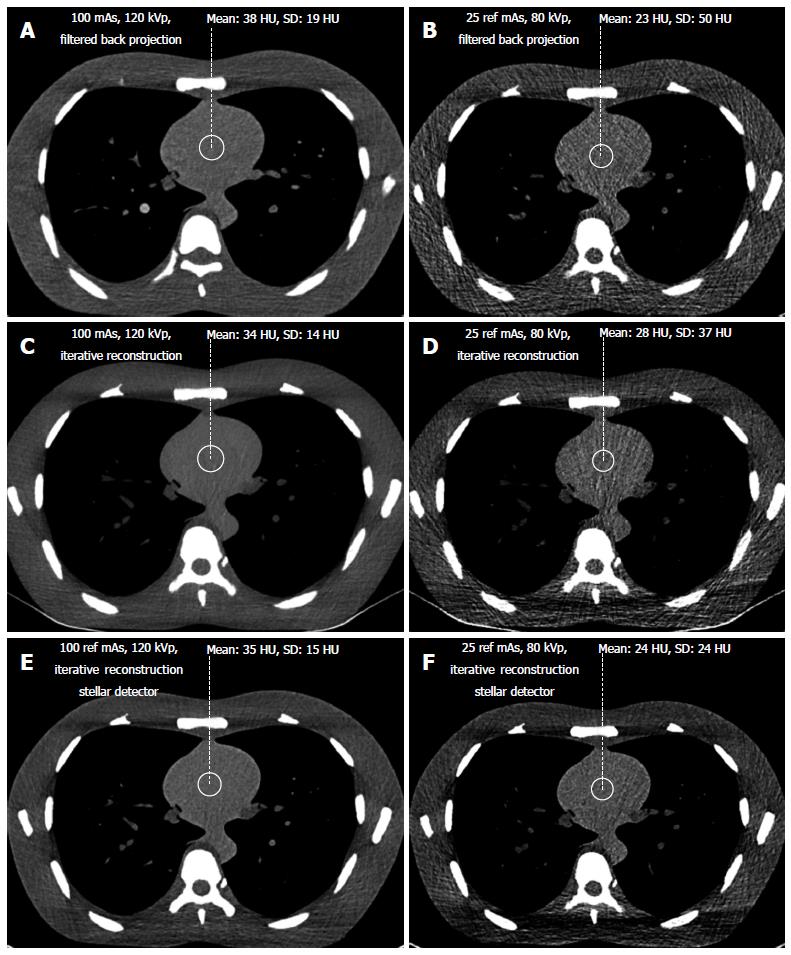
Figure 2 Soft tissue imaging using filtered back projection (A, B), iterative reconstruction (C, D, E, F) and the Stellar detector (E, F).
At standard dose levels (A, C, E), image quality decreased from left to right. At the lowest dose level (B, D, F), the difference in noise increased. The image quality of the low dose image with the Stellar detector (F) was close to the image quality of that for a standard dose with a filtered back projection (A).
- Citation: Christe A, Heverhagen J, Ozdoba C, Weisstanner C, Ulzheimer S, Ebner L. CT dose and image quality in the last three scanner generations. World J Radiol 2013; 5(11): 421-429
- URL: https://www.wjgnet.com/1949-8470/full/v5/i11/421.htm
- DOI: https://dx.doi.org/10.4329/wjr.v5.i11.421


