Copyright
©2013 Baishideng Publishing Group Co.
World J Radiol. Oct 28, 2013; 5(10): 356-371
Published online Oct 28, 2013. doi: 10.4329/wjr.v5.i10.356
Published online Oct 28, 2013. doi: 10.4329/wjr.v5.i10.356
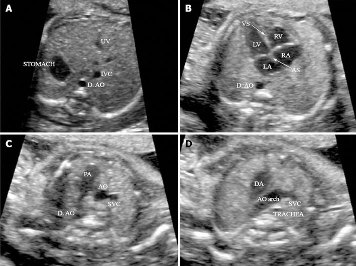
Figure 4 Four routine axial views of heart and great vessels.
A: Transverse view of the superior abdomen: the stomach on the fetal left side; the descending aorta (D. AO) to the left side and inferior vena cava (IVC) to right side of the spine, respectively; B: Four-chamber view: in a normal fetal heart, approximately equal size of the right and left chambers, intact intact ventricular septum (VS) and normal offset of the two atrioventricular valve; C: Three-vessel view: pulmonary artery (PA), aorta (AO) and superior vena cava (SVC) in the correct position and alignment; PA, to the left, is the largest of the three and the most anterior, whereas the SVC is the smallest and most posterior; D: Transverse view of the aortic arch: in the normal heart, both the AO arch and the ductal arch (DA) are located to the left of the trachea, in a ‘V’-shaped configuration. (Adapted from ISUOG Practice Guidelines[115]). UV: umbilical vein; RV: right ventricle; LV: left ventricle; LA: left atrium; RA: right atrium; AS: Atrial septum.
- Citation: Renna MD, Pisani P, Conversano F, Perrone E, Casciaro E, Renzo GCD, Paola MD, Perrone A, Casciaro S. Sonographic markers for early diagnosis of fetal malformations. World J Radiol 2013; 5(10): 356-371
- URL: https://www.wjgnet.com/1949-8470/full/v5/i10/356.htm
- DOI: https://dx.doi.org/10.4329/wjr.v5.i10.356


