Copyright
©2012 Baishideng Publishing Group Co.
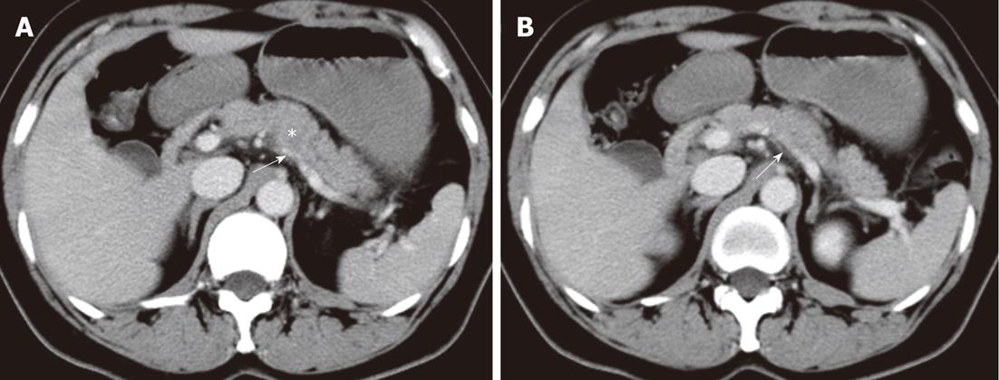
Figure 3 Patterns of extrapancreatic neural invasion by pancreatic carcinoma visualized by computed tomography in a 56-year-old female with pancreatic carcinoma in the pancreas body.
Contrast-enhanced computed tomography images (A, B) show the splenic plexus invasion (arrows) by the tumor (asterisk) in the body of pancreas. The appearance of plexus invasion is streaky structure.
- Citation: Zuo HD, Tang W, Zhang XM, Zhao QH, Xiao B. CT and MR imaging patterns for pancreatic carcinoma invading the extrapancreatic neural plexus (Part II): Imaging of pancreatic carcinoma nerve invasion. World J Radiol 2012; 4(1): 13-20
- URL: https://www.wjgnet.com/1949-8470/full/v4/i1/13.htm
- DOI: https://dx.doi.org/10.4329/wjr.v4.i1.13


