Copyright
©2012 Baishideng Publishing Group Co.
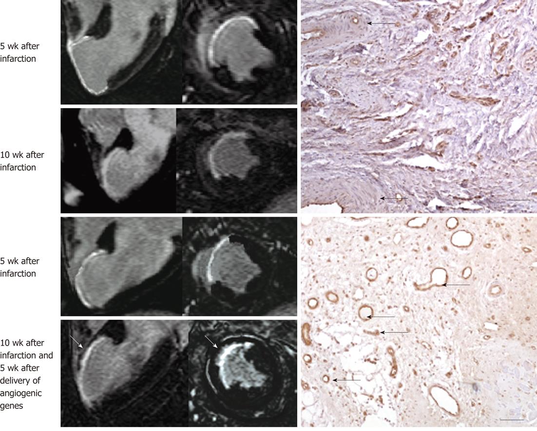
Figure 8 Gadolinium-enhanced magnetic resonance long-axis (left column) and short-axis (right column) images illustrate the extent of myocardial infarct (hyperenhanced myocardium) at 5 wk after infarction and 5 wk after injection of saline (top block, 4 magnetic resonance images) and angiogenic gene (bottom block, 4 magnetic resonance images).
Gadolinium-enhanced magnetic resonance (MR) images delineated myocardial infarct and showed substantial reduction in infarct extent and transmurality 5 wk after treatment (bottom block, bottom row, white arrows) compared with control animal (top block, bottom row). The angiogenic gene was delivered transendocardially under MR-guidance as shown in Figure 2. The histopathologic sections (right) show very few blood vessels in control animal (top right, black arrows) and the formation of abundant new blood vessels in gene treated animal (bottom right, black arrows).
- Citation: Saeed M, Wilson M. Value of MR contrast media in image-guided body interventions. World J Radiol 2012; 4(1): 1-12
- URL: https://www.wjgnet.com/1949-8470/full/v4/i1/1.htm
- DOI: https://dx.doi.org/10.4329/wjr.v4.i1.1


