
















©2011 Baishideng Publishing Group Co.
World J Radiol. Sep 28, 2011; 3(9): 224-232
Published online Sep 28, 2011. doi: 10.4329/wjr.v3.i9.224
Published online Sep 28, 2011. doi: 10.4329/wjr.v3.i9.224
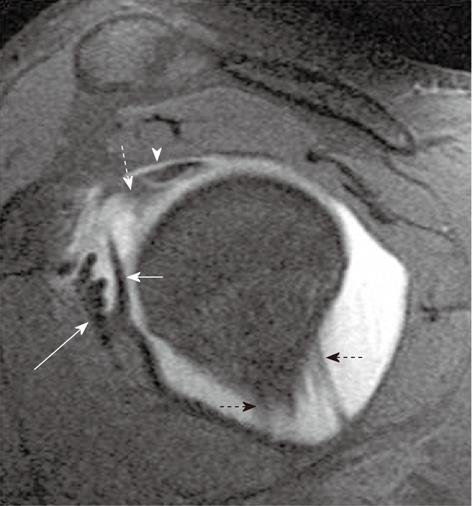
Figure 4 Oblique sagittal T1-weighted fat-saturated magnetic resonance arthrogram image shows normal superior glenohumeral ligament (white dashed arrow), inferior to the intra-articular long head of biceps tendon (arrowhead).
The middle glenohumeral ligament is seen as a long hypointense band (short straight arrow) medial to the subscapularis tendon (long straight arrow). Anterior and posterior bands of inferior glenohumeral ligament are shown with black dashed arrows.
- Citation: Jana M, Gamanagatti S. Magnetic resonance imaging in glenohumeral instability. World J Radiol 2011; 3(9): 224-232
- URL: https://www.wjgnet.com/1949-8470/full/v3/i9/224.htm
- DOI: https://dx.doi.org/10.4329/wjr.v3.i9.224