Copyright
©2011 Baishideng Publishing Group Co.
World J Radiol. Dec 28, 2011; 3(12): 289-297
Published online Dec 28, 2011. doi: 10.4329/wjr.v3.i12.289
Published online Dec 28, 2011. doi: 10.4329/wjr.v3.i12.289
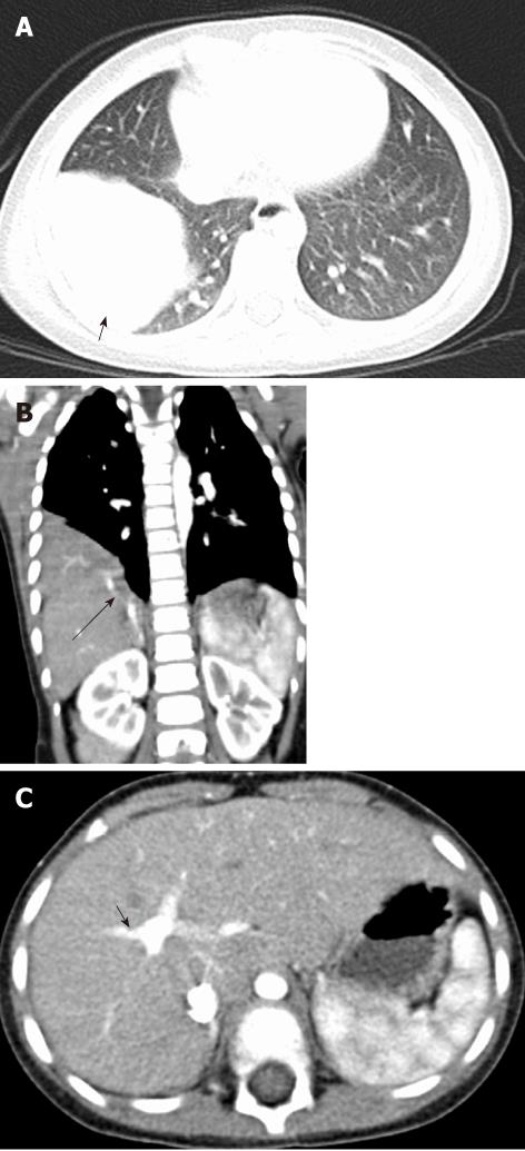
Figure 15 Sequestration of lung segment- Extralobar type.
A: Axial image in the lung window shows a homogenous pulmonary lesion in the location of the right lower lobe lateral basal segment; B, C: Coronal multiplanar reformatted images (B) and axial image (C) at the level of portal vein bifurcation in the mediastinal window show anomalous venous drainage (long arrow) into the right branch of the portal vein (short arrow).
- Citation: Sundarakumar DK, Bhalla AS, Sharma R, Gupta AK, Kabra SK, Jagia P. Multidetector computed tomography imaging of congenital anomalies of major airways: A pictorial essay. World J Radiol 2011; 3(12): 289-297
- URL: https://www.wjgnet.com/1949-8470/full/v3/i12/289.htm
- DOI: https://dx.doi.org/10.4329/wjr.v3.i12.289


