Copyright
©2011 Baishideng Publishing Group Co.
World J Radiol. Dec 28, 2011; 3(12): 289-297
Published online Dec 28, 2011. doi: 10.4329/wjr.v3.i12.289
Published online Dec 28, 2011. doi: 10.4329/wjr.v3.i12.289
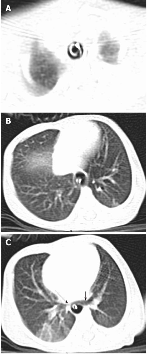
Figure 1 Tracheal agenesis type-III.
A, B: Axial images in the lung window show the presence of dilated esophagus and tube in situ. There is no separate lumen for trachea; C: Caudally, right (long arrow) and left main bronchi (short arrow) arise independently from the thoracic esophagus.
- Citation: Sundarakumar DK, Bhalla AS, Sharma R, Gupta AK, Kabra SK, Jagia P. Multidetector computed tomography imaging of congenital anomalies of major airways: A pictorial essay. World J Radiol 2011; 3(12): 289-297
- URL: https://www.wjgnet.com/1949-8470/full/v3/i12/289.htm
- DOI: https://dx.doi.org/10.4329/wjr.v3.i12.289


