Copyright
©2010 Baishideng Publishing Group Co.
World J Radiol. Sep 28, 2010; 2(9): 358-367
Published online Sep 28, 2010. doi: 10.4329/wjr.v2.i9.358
Published online Sep 28, 2010. doi: 10.4329/wjr.v2.i9.358
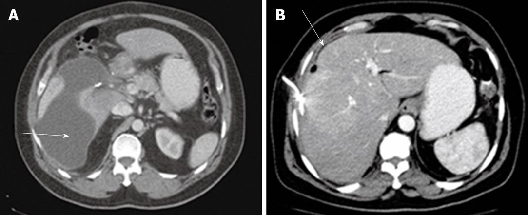
Figure 8 A case of post cholecystectomy biloma.
A: Abdominal computed tomography (CT) shows a large subhepatic biloma (arrow); B: CT-guided drainage of the biloma shows marked regression of the volume of the collection (arrow).
- Citation: Donkol RH, Latif NA, Moghazy K. Percutaneous imaging-guided interventions for acute biliary disorders in high surgical risk patients. World J Radiol 2010; 2(9): 358-367
- URL: https://www.wjgnet.com/1949-8470/full/v2/i9/358.htm
- DOI: https://dx.doi.org/10.4329/wjr.v2.i9.358


