Copyright
©2010 Baishideng Publishing Group Co.
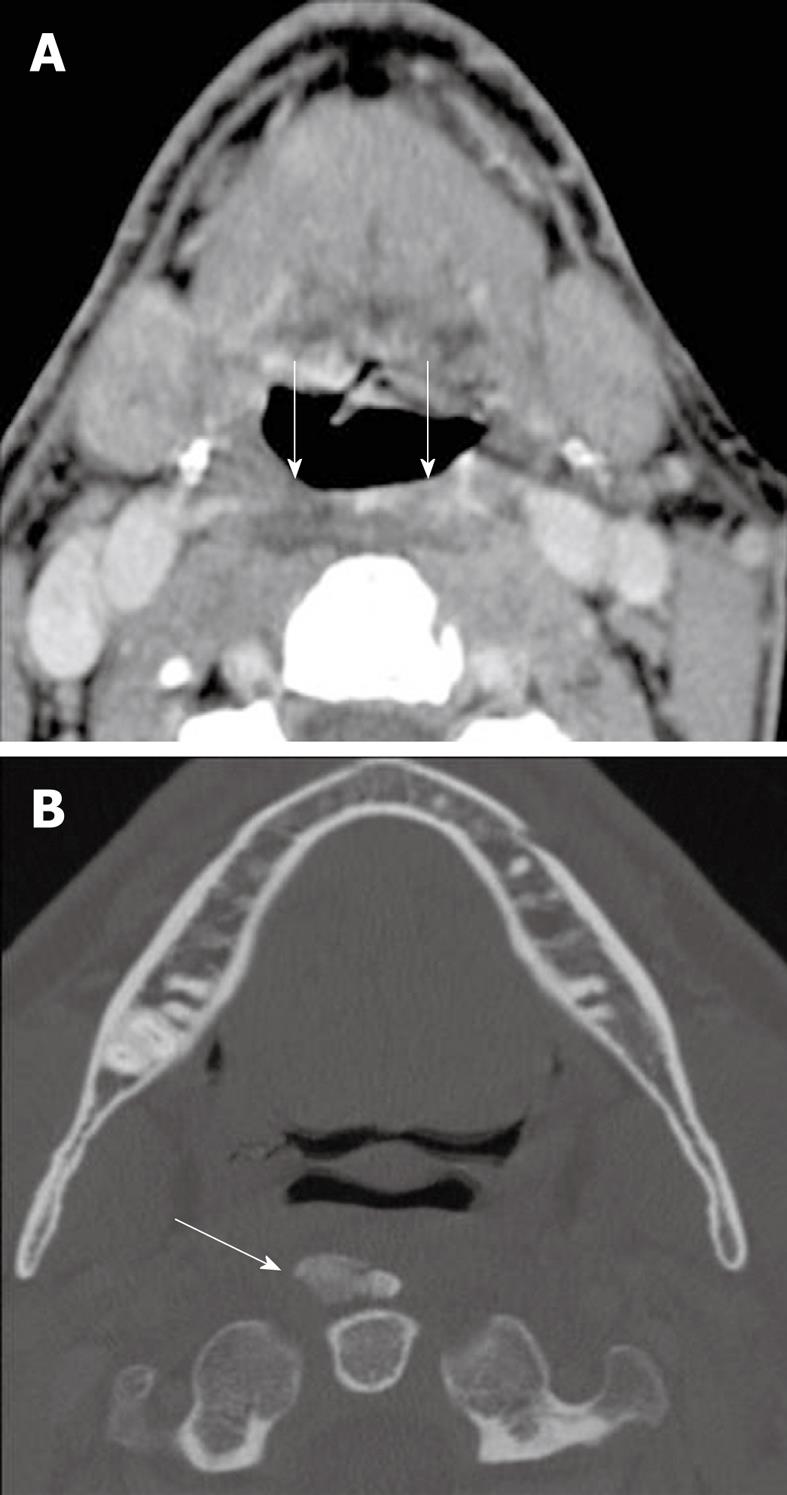
Figure 12 Calcific tendinitis.
A: Axial contrast enhanced CT of the neck demonstrates retropharyngeal space edema (arrows); B: The bone algorithm shows an ossific mass anterior to the dens confirming that the edema in the retropharyngeal space is due to calcific tendinitis (arrow).
- Citation: Bou-Assaly W, Mckellop J, Mukherji S. Computed tomography imaging of acute neck inflammatory processes. World J Radiol 2010; 2(3): 91-96
- URL: https://www.wjgnet.com/1949-8470/full/v2/i3/91.htm
- DOI: https://dx.doi.org/10.4329/wjr.v2.i3.91


