Copyright
©2010 Baishideng Publishing Group Co.
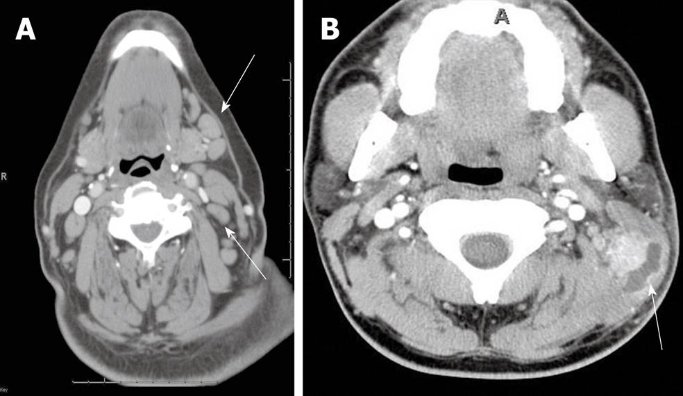
Figure 1 Cervical and suppurative adenitis.
A: Axial contrast-enhanced computed tomography (CT) shows homogenous enlargement of multiple enlarged to borderline sized lymph nodes (arrows), in a patient with neck pain consistent with cervical adenitis; B: Axial contrast-enhanced CT shows a suppurative cervical lymph node (arrow) with surrounding soft tissue edema.
- Citation: Bou-Assaly W, Mckellop J, Mukherji S. Computed tomography imaging of acute neck inflammatory processes. World J Radiol 2010; 2(3): 91-96
- URL: https://www.wjgnet.com/1949-8470/full/v2/i3/91.htm
- DOI: https://dx.doi.org/10.4329/wjr.v2.i3.91


