Copyright
©2010 Baishideng Publishing Group Co.
World J Radiol. Dec 28, 2010; 2(12): 468-471
Published online Dec 28, 2010. doi: 10.4329/wjr.v2.i12.468
Published online Dec 28, 2010. doi: 10.4329/wjr.v2.i12.468
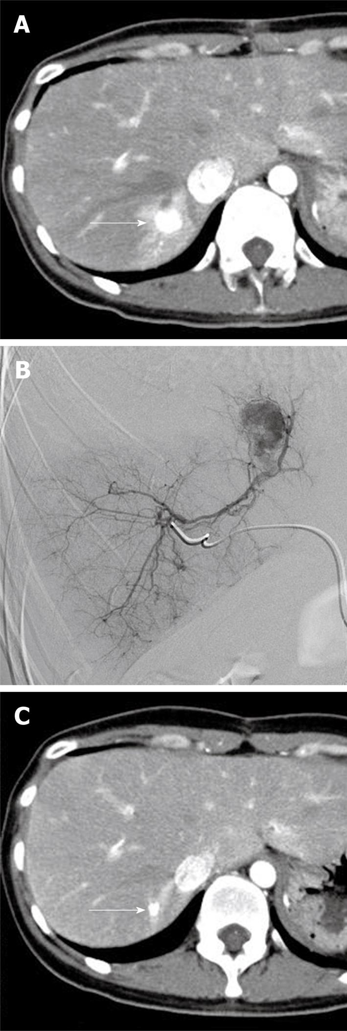
Figure 2 Images obtained in the case of a 35-year-old woman with a solitary neuroendocrine metastasis of the liver.
A: Arterial phase image of contrast-enhanced computed tomography (CT) before intervention shows a hyperdense metastatic tumor in the right posterior segment surrounded by arterioportal shunting (arrow); B: Selective angiogram from the right posterior arterial segment delineates a single hypervascular metastatic tumor with arterioportal shunting; C: Arterial phase image of contrast-enhanced CT at 3 mo after chemoembolization with miriplatin-iodized oil suspension demonstrates a significant reduction in tumor size with compact accumulation of iodized oil accompanied by surrounding arterioportal shunting (arrow).
- Citation: Iwazawa J, Ohue S, Yasumasa K, Mitani T. Transarterial chemoembolization with miriplatin-lipiodol emulsion for neuroendocrine metastases of the liver. World J Radiol 2010; 2(12): 468-471
- URL: https://www.wjgnet.com/1949-8470/full/v2/i12/468.htm
- DOI: https://dx.doi.org/10.4329/wjr.v2.i12.468


