Copyright
©2010 Baishideng Publishing Group Co.
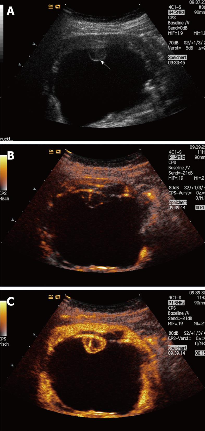
Figure 5 Cystic renal lesion with a small RCC (12 mm × 10 mm) not recognized by CT which has been histologically proven by surgery.
B-mode US showed a nodularity inside the cyst (A); CEUS revealed contrast enhancement of the small lesion (B, C)[134].
- Citation: Ignee A, Straub B, Schuessler G, Dietrich CF. Contrast enhanced ultrasound of renal masses. World J Radiol 2010; 2(1): 15-31
- URL: https://www.wjgnet.com/1949-8470/full/v2/i1/15.htm
- DOI: https://dx.doi.org/10.4329/wjr.v2.i1.15


