Copyright
©2010 Baishideng Publishing Group Co.
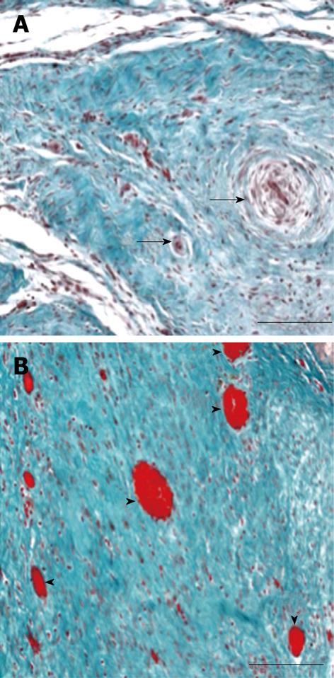
Figure 5 Micrographs of representative infarcts in control and VEGF-gene treated swine.
A: The infarct in the control animal shows no appreciable angiogenesis and residual blood vessels have been remodeled, as shown by the thick vascular wall and small lumen (black arrows). B: VEGF-gene treated animal contains numerous blood vessels (arrowheads) in linear array representing injection track (calibration bar = 200 μm)[60].
- Citation: Saeed M, Hetts S, Wilson M. Reperfusion injury components and manifestations determined by cardiovascular MR and MDCT imaging. World J Radiol 2010; 2(1): 1-14
- URL: https://www.wjgnet.com/1949-8470/full/v2/i1/1.htm
- DOI: https://dx.doi.org/10.4329/wjr.v2.i1.1


