
















Copyright: ©Author(s) 2026.
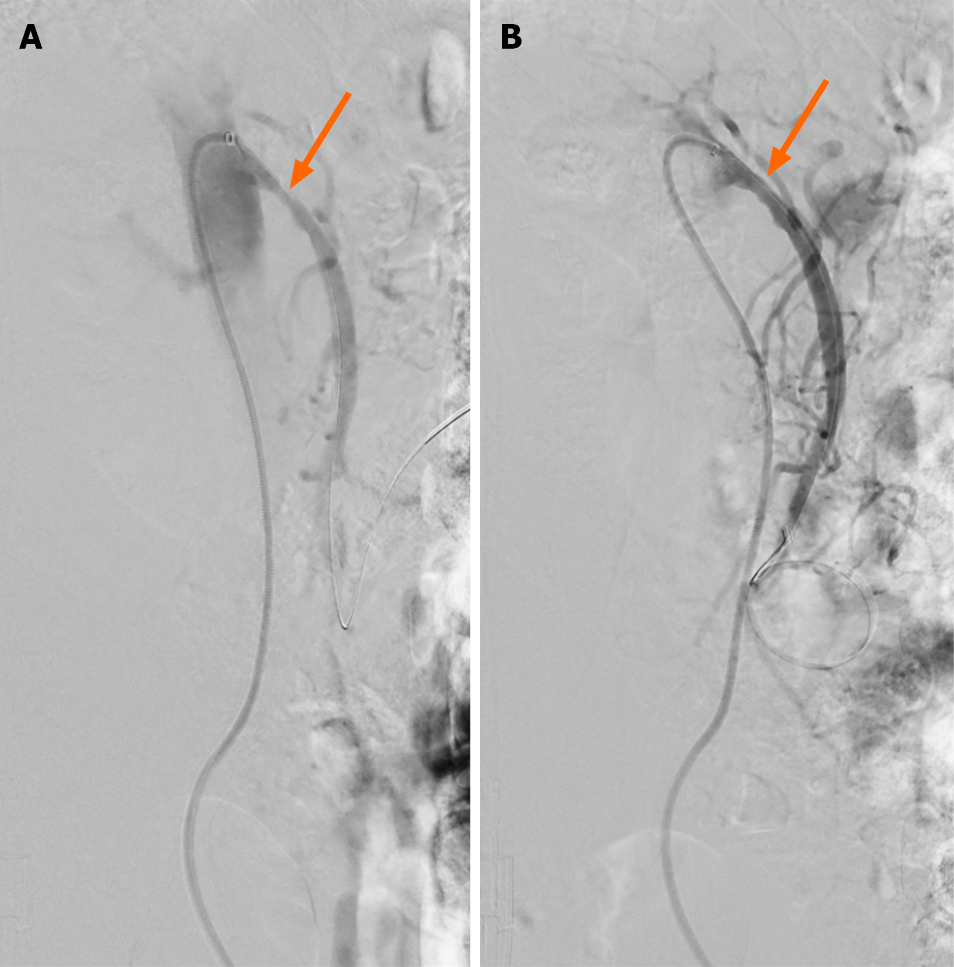
Figure 7 Angiographic result of covered stent placement for proximal superior mesenteric artery stenosis.
A: Lateral digital subtraction angiographic image via a 6-Fr sheath at the ostium of the superior mesenteric artery demonstrating severe proximal stenosis (arrow); B: Corresponding image following covered stent placement demonstrating improved flow across the treated segment, with residual stenosis of approximately 20% secondary to extensive calcified atherosclerosis (arrow). Same patient as Figures 5 and 6.
- Citation: Keady C, O’Malley CM, Mohamed SA, Bell J, Crotty C, Patton A, O’Regan D, Keeling A, McGrath A, Given M, Mulholland D, Sheehan M. Endovascular management of acute and chronic arterial mesenteric ischaemia: A narrative review of evidence informing interventional radiology treatment. World J Radiol 2026; 18(10): 125052
- URL: https://www.wjgnet.com/1949-8470/full/v18/i10/125052.htm
- DOI: https://dx.doi.org/10.4329/wjr.125052