
















Copyright: ©Author(s) 2026.
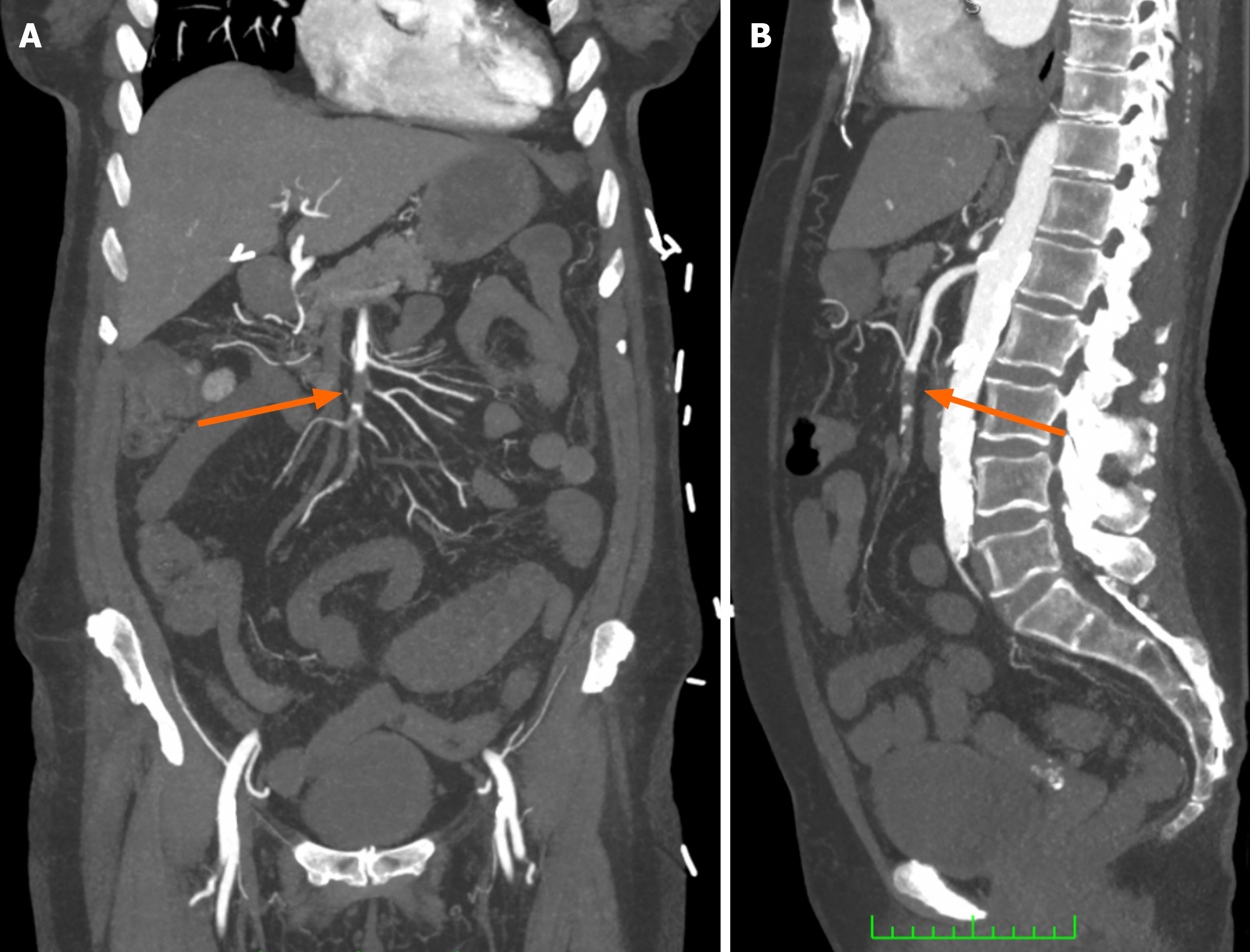
Figure 1 Acute superior mesenteric artery occlusion on computed tomography angiography.
A: Coronal arterial phase image demonstrating an extensive intraluminal filling defect within the middle segment of the superior mesenteric artery (arrow); B: Sagittal arterial phase image demonstrating the same filling defect (arrow). Images from a 65-year-old female presenting with acute embolic mesenteric ischaemia; same patient as Figures 2, 3, and 4.
- Citation: Keady C, O’Malley CM, Mohamed SA, Bell J, Crotty C, Patton A, O’Regan D, Keeling A, McGrath A, Given M, Mulholland D, Sheehan M. Endovascular management of acute and chronic arterial mesenteric ischaemia: A narrative review of evidence informing interventional radiology treatment. World J Radiol 2026; 18(10): 125052
- URL: https://www.wjgnet.com/1949-8470/full/v18/i10/125052.htm
- DOI: https://dx.doi.org/10.4329/wjr.125052