Copyright
©The Author(s) 2025.
World J Radiol. Aug 28, 2025; 17(8): 110868
Published online Aug 28, 2025. doi: 10.4329/wjr.v17.i8.110868
Published online Aug 28, 2025. doi: 10.4329/wjr.v17.i8.110868
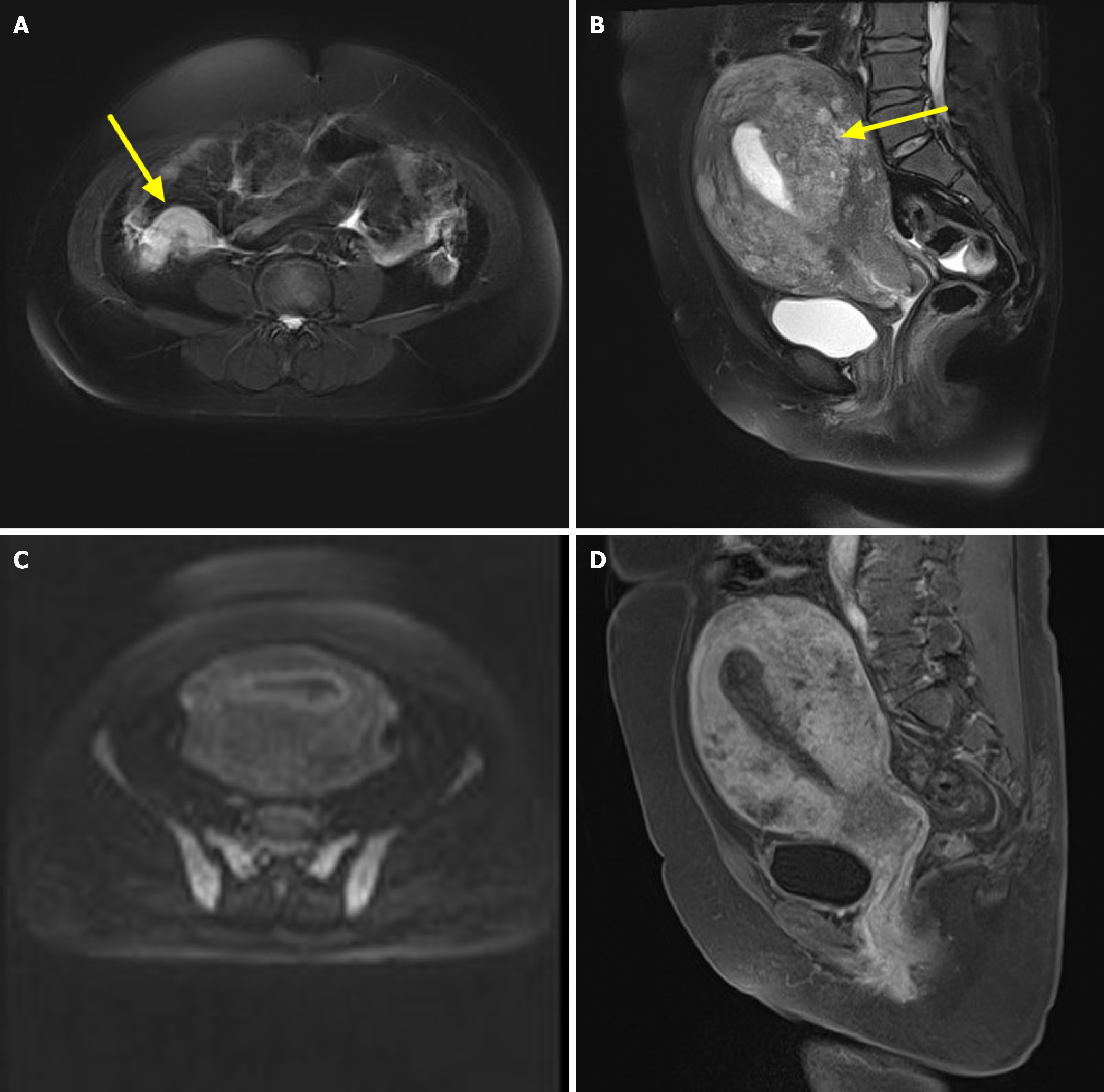
Figure 2 Magnetic resonance imaging features of appendix and uterus.
A: T2-weighted imaging (T2WI) transverse view: Coarsening appendix (arrowhead), T2WI high signal, and signals are relatively uniform; B: T2WI sagittal view, obvious thickening mesometrium (arrowhead), and ambiguous display of bonding zone; C: Diffusion-weighted imaging, the enlarged uterus presents slightly high signals; D: T1-weighted imaging enhanced sagittal view, strengthening mesometrium and relatively complete serosa.
- Citation: Liu JM, Li Z, Qi LH, Chu BL, Deng ZX, Tang FY. Imaging features of appendiceal signet ring cell carcinoma with uterine implantation: A case report. World J Radiol 2025; 17(8): 110868
- URL: https://www.wjgnet.com/1949-8470/full/v17/i8/110868.htm
- DOI: https://dx.doi.org/10.4329/wjr.v17.i8.110868


