Copyright
©The Author(s) 2025.
World J Radiol. Aug 28, 2025; 17(8): 110868
Published online Aug 28, 2025. doi: 10.4329/wjr.v17.i8.110868
Published online Aug 28, 2025. doi: 10.4329/wjr.v17.i8.110868
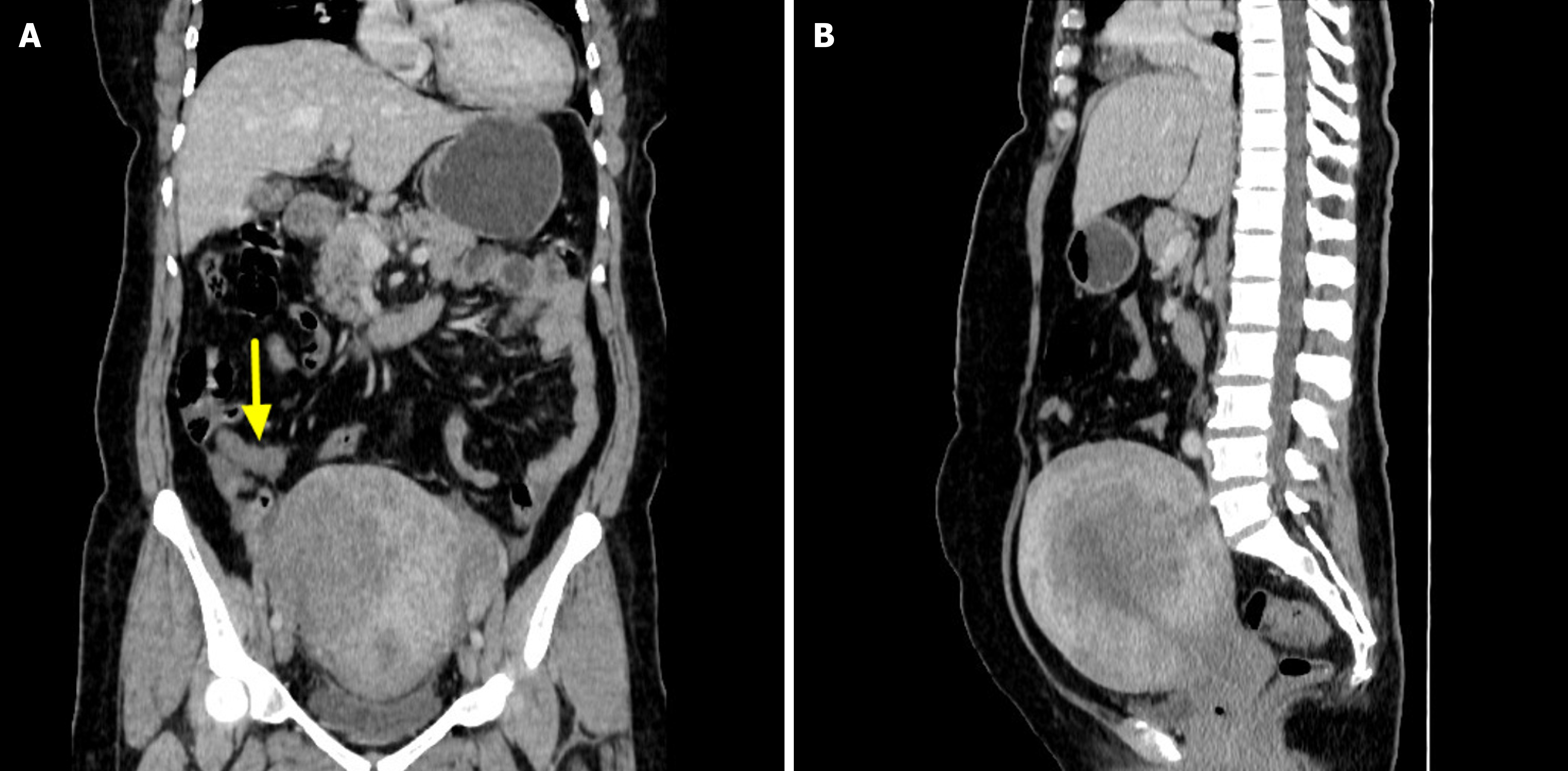
Figure 1 Computed tomography features of the appendix and uterus.
A: Computed tomography (CT) enhanced coronal view: Coarsening appendix (arrowhead), strengthening after enhancement, clear boundary between focus and surrounding tissues; B: CT enhanced sagittal view: Obvious uterus enlargement, obvious muscular.
- Citation: Liu JM, Li Z, Qi LH, Chu BL, Deng ZX, Tang FY. Imaging features of appendiceal signet ring cell carcinoma with uterine implantation: A case report. World J Radiol 2025; 17(8): 110868
- URL: https://www.wjgnet.com/1949-8470/full/v17/i8/110868.htm
- DOI: https://dx.doi.org/10.4329/wjr.v17.i8.110868


