
















©The Author(s) 2025.
World J Radiol. Aug 28, 2025; 17(8): 110407
Published online Aug 28, 2025. doi: 10.4329/wjr.v17.i8.110407
Published online Aug 28, 2025. doi: 10.4329/wjr.v17.i8.110407
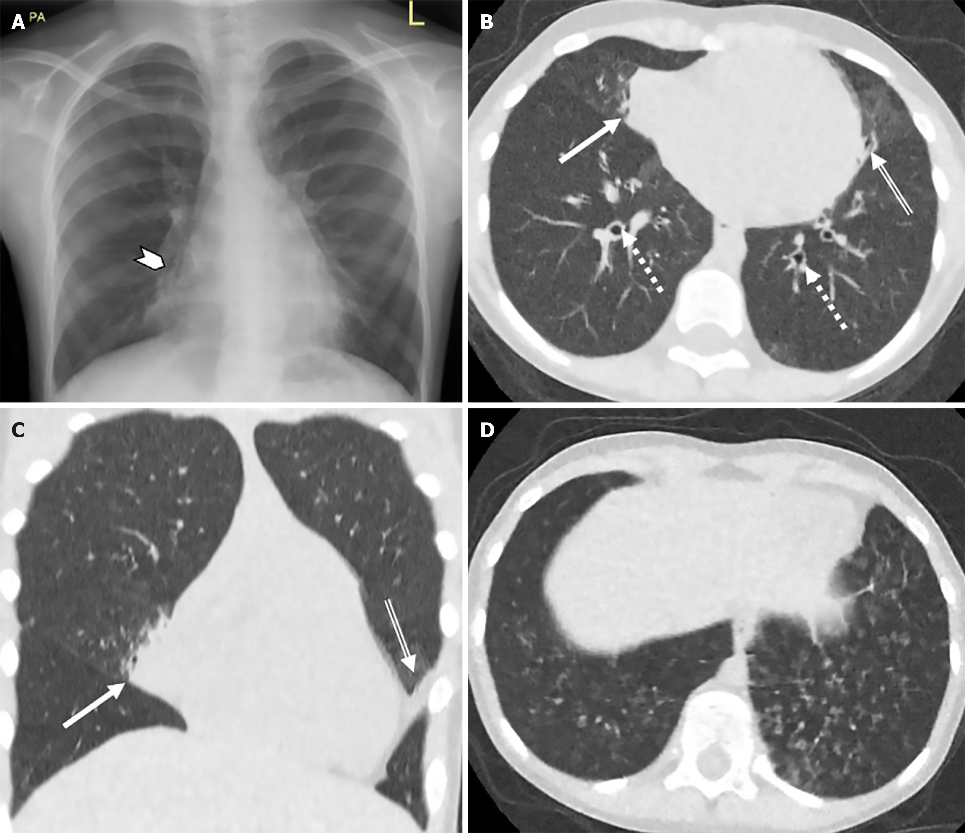
Figure 1 Chest radiography and ultra-low dose computed tomography chest assessment of structural lung pathology.
A: Chest radiography; B: Axial ultra-low dose computed tomography (ULDCT); C: Coronal ULDCT; D: Axial ULDCT with lung windows of a 7-year-old female with dense medial middle lobe consolidation (solid arrow & arrowhead), bilateral bronchiectasis (dashed arrow), lingular atelectasis (double line arrow) and basal centrilobular ground-glass nodules (D). These representative images demonstrate the superiority of ULDCT in assessment of structural changes relating to primary ciliary dyskinesia in comparison to plain radiography.
- Citation: Waldron MG, O'Regan PW, Lane M, Shet SS, Kakish E, Moloney F, Moore N, Murphy MJ, Beagan L, Plant BJ, Mullane D, Ni Chroinin M, Ryan DJ, O'Regan K, Power SP, Maher MM. Ultra-low dose computed tomography chest vs chest radiography in paediatric primary ciliary dyskinesia: A prospective study. World J Radiol 2025; 17(8): 110407
- URL: https://www.wjgnet.com/1949-8470/full/v17/i8/110407.htm
- DOI: https://dx.doi.org/10.4329/wjr.v17.i8.110407