Copyright
©The Author(s) 2025.
World J Radiol. Aug 28, 2025; 17(8): 110307
Published online Aug 28, 2025. doi: 10.4329/wjr.v17.i8.110307
Published online Aug 28, 2025. doi: 10.4329/wjr.v17.i8.110307
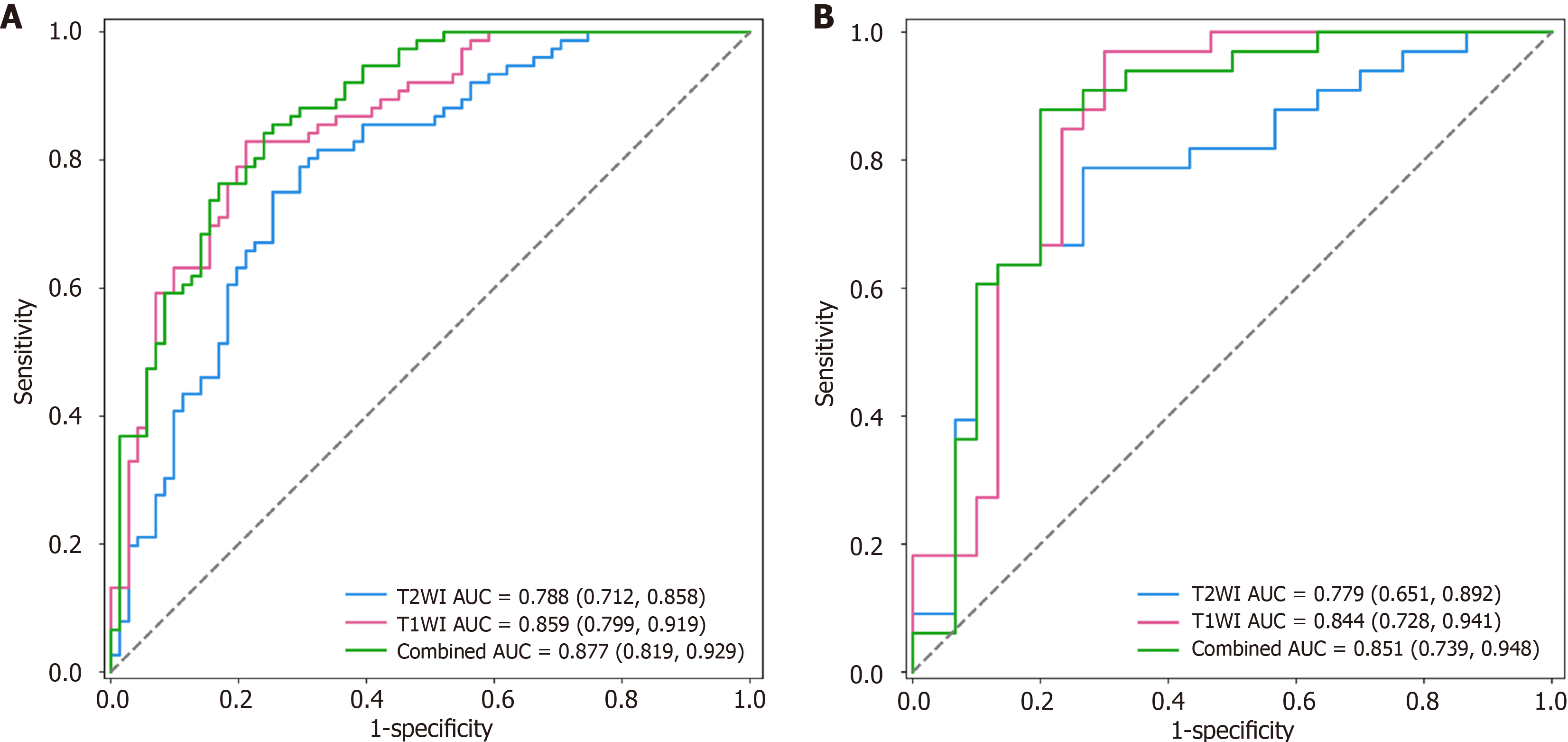
Figure 2 Receiver operating characteristic curves of the radiomics signature’s discriminative performance in esophageal cancer staging in the primary and validation cohorts.
A: Primary cohorts; B: Validation cohorts. The combined radiomics signature demonstrated superior area under the curve (AUC) in both the primary (AUC: 0.877) and validation cohorts (AUC: 0.851), outperforming single-sequence models. AUC: Area under the curve; T1WI: T1-weighted imaging; T2WI: T2-weighted imaging.
- Citation: Yang RH, Lin ZP, Dong T, Fan WX, Qin HD, Jiang GH, Dai HY. Magnetic resonance imaging-based radiomics signature for predicting preoperative staging of esophageal cancer. World J Radiol 2025; 17(8): 110307
- URL: https://www.wjgnet.com/1949-8470/full/v17/i8/110307.htm
- DOI: https://dx.doi.org/10.4329/wjr.v17.i8.110307


