Copyright
©The Author(s) 2025.
World J Radiol. Jun 28, 2025; 17(6): 107205
Published online Jun 28, 2025. doi: 10.4329/wjr.v17.i6.107205
Published online Jun 28, 2025. doi: 10.4329/wjr.v17.i6.107205
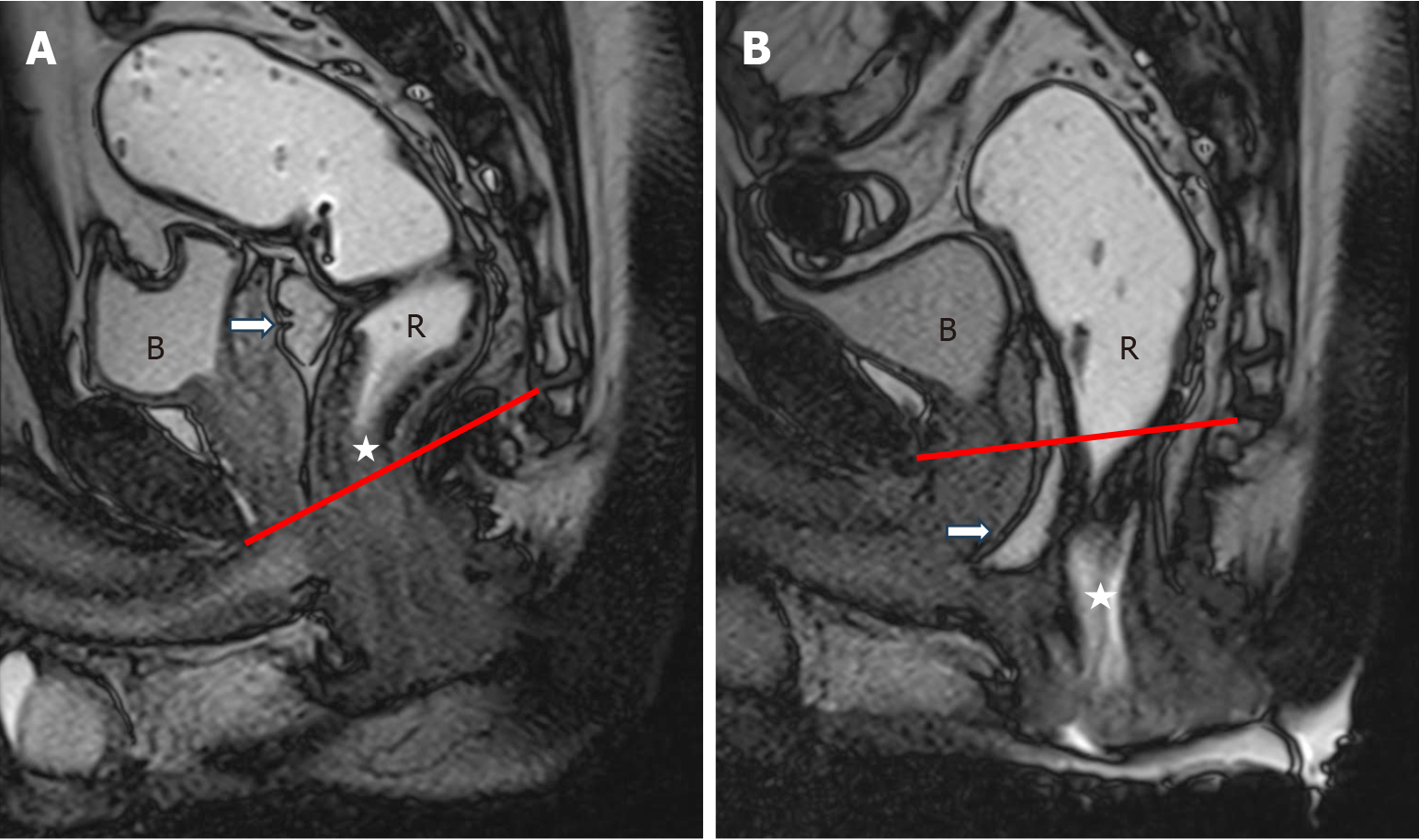
Figure 7 Peritoneocele.
A: Sagittal true fast imaging with steady-state free precession image at rest shows the normal position of cul-de-sac (white arrow) above pubococcygeal line (PCL); B: During the defecation phase there is caudal descent of the peritoneum (peritoneocele) (white arrow) below the PCL. Note descent of anorectal junction (white star).
- Citation: Parry AH, Rehaman B, Bhat SA, Wani AH, Jehangir M, Baba AA. Role of magnetic resonance defecography in the assessment of obstructed defecation syndrome. World J Radiol 2025; 17(6): 107205
- URL: https://www.wjgnet.com/1949-8470/full/v17/i6/107205.htm
- DOI: https://dx.doi.org/10.4329/wjr.v17.i6.107205


