Copyright
©The Author(s) 2025.
World J Radiol. Jun 28, 2025; 17(6): 107205
Published online Jun 28, 2025. doi: 10.4329/wjr.v17.i6.107205
Published online Jun 28, 2025. doi: 10.4329/wjr.v17.i6.107205
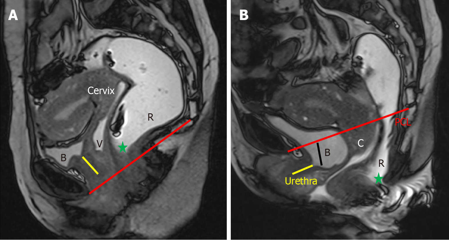
Figure 5 Urethral hypermobility and cystocele.
A: Sagittal true fast imaging with steady-state free precession (TRUFI) at rest, which shows minimal posterior angulation of the urethra (yellow line), approximately 5° relative to the vertical axis of the pubis; B: In the sagittal TRUFI image obtained during the defecation phase, the urethral axis rotates posteriorly to 162° and descends below the pubic symphysis, indicating urethral hypermobility. This is accompanied by a descent of the bladder neck below the pubococcygeal line, consistent with cystocele.
- Citation: Parry AH, Rehaman B, Bhat SA, Wani AH, Jehangir M, Baba AA. Role of magnetic resonance defecography in the assessment of obstructed defecation syndrome. World J Radiol 2025; 17(6): 107205
- URL: https://www.wjgnet.com/1949-8470/full/v17/i6/107205.htm
- DOI: https://dx.doi.org/10.4329/wjr.v17.i6.107205


