Copyright
©The Author(s) 2025.
World J Radiol. Jun 28, 2025; 17(6): 107205
Published online Jun 28, 2025. doi: 10.4329/wjr.v17.i6.107205
Published online Jun 28, 2025. doi: 10.4329/wjr.v17.i6.107205
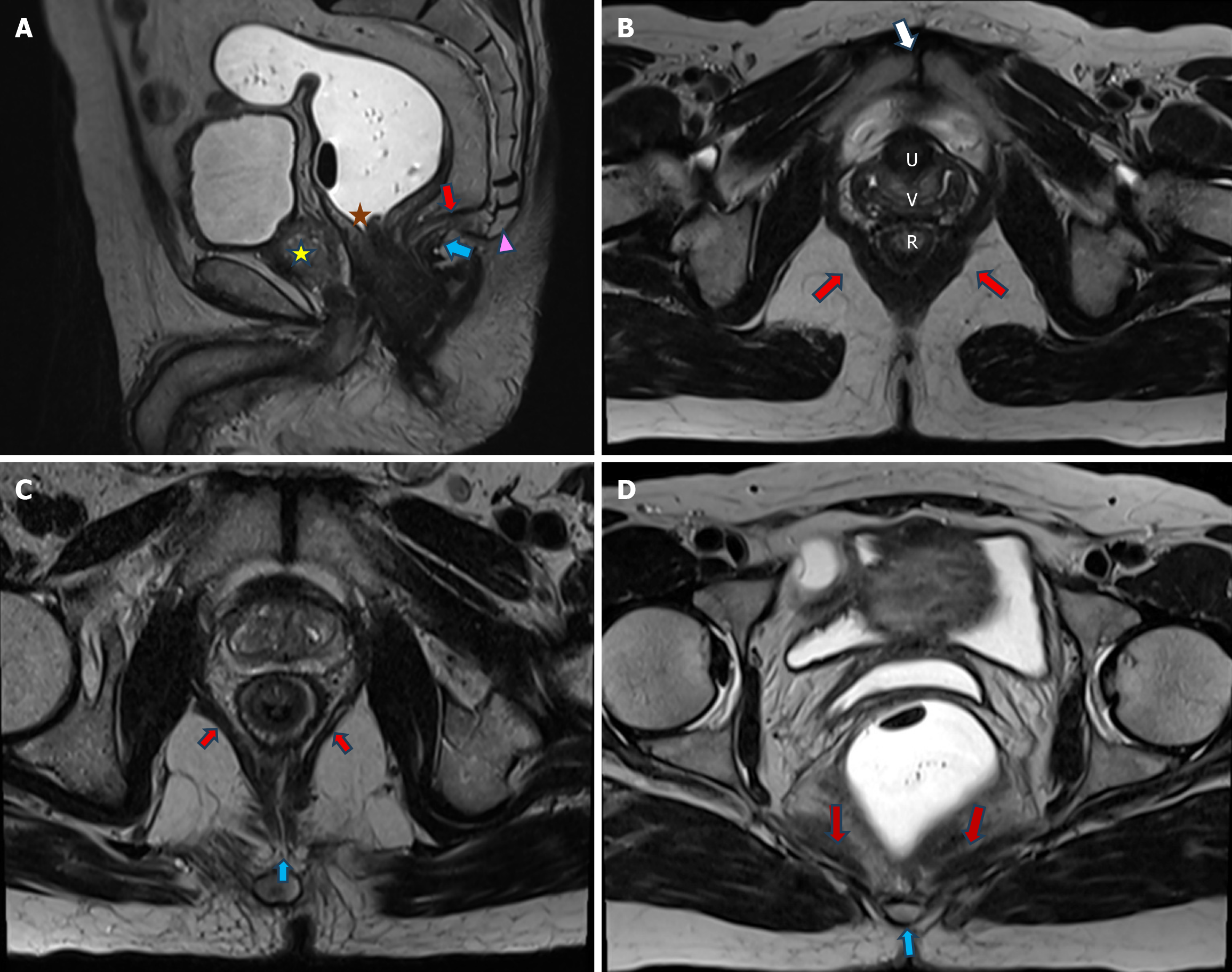
Figure 2 Normal supporting structures of pelvic floor.
A: T2-weighted image in sagittal plane shows the posterior aspect of puborectalis muscle (blue arrow) just behind the anorectal junction (orange star). Levator plate (red arrow) extends from the coccyx (pink arrowhead) to the anorectal junction (orange star). Note the normal vertical orientation of the urethra (yellow star) behind pubic bone. Bladder (B) and rectum (R) are distended; B: Axial T2-weighted image shows the sling of puborectalis muscle (red arrows) attached to the pubis (white arrow) anteriorly and encircling around the anorectal junction (R) posteriorly; C: Axial T2-weighted image cephalad to (B) shows pubococcygeus muscle (red arrows) coursing from the pubis anteriorly to the levator plate (blue arrow) posteriorly; D: Axial T2-weighted image cephalad to (C) shows iliococcygeus muscle (red arrows) extending from the obturator fascia laterally to the coccyx (blue arrow) posteriorly.
- Citation: Parry AH, Rehaman B, Bhat SA, Wani AH, Jehangir M, Baba AA. Role of magnetic resonance defecography in the assessment of obstructed defecation syndrome. World J Radiol 2025; 17(6): 107205
- URL: https://www.wjgnet.com/1949-8470/full/v17/i6/107205.htm
- DOI: https://dx.doi.org/10.4329/wjr.v17.i6.107205


