Copyright
©The Author(s) 2025.
World J Radiol. Jun 28, 2025; 17(6): 107205
Published online Jun 28, 2025. doi: 10.4329/wjr.v17.i6.107205
Published online Jun 28, 2025. doi: 10.4329/wjr.v17.i6.107205
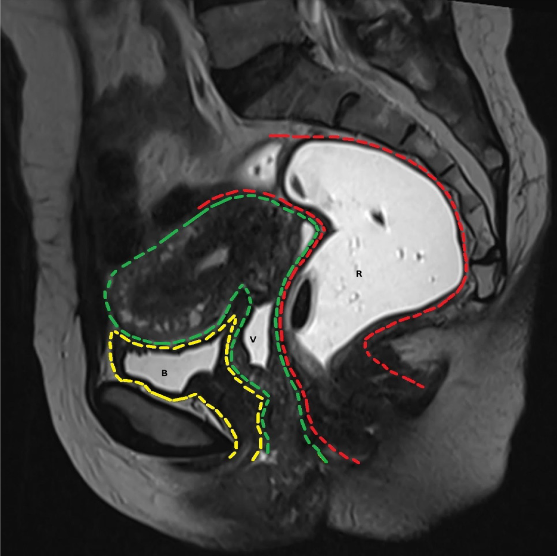
Figure 1 T2-weighted image in sagittal plane shows the three compartments of pelvis.
The anterior compartment (yellow) contains the urinary bladder (B) and urethra, middle compartment (green) contains the uterus, cervix and vagina (V) and the posterior compartment (red) contains the rectum (R) and anal canal.
- Citation: Parry AH, Rehaman B, Bhat SA, Wani AH, Jehangir M, Baba AA. Role of magnetic resonance defecography in the assessment of obstructed defecation syndrome. World J Radiol 2025; 17(6): 107205
- URL: https://www.wjgnet.com/1949-8470/full/v17/i6/107205.htm
- DOI: https://dx.doi.org/10.4329/wjr.v17.i6.107205


