Copyright
©The Author(s) 2025.
World J Radiol. Jun 28, 2025; 17(6): 107164
Published online Jun 28, 2025. doi: 10.4329/wjr.v17.i6.107164
Published online Jun 28, 2025. doi: 10.4329/wjr.v17.i6.107164
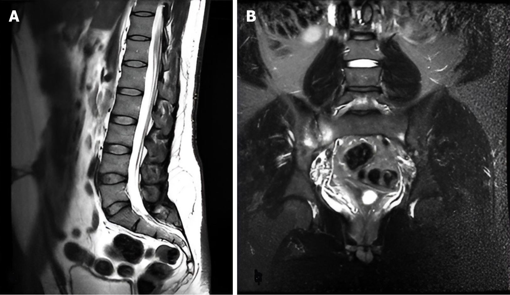
Figure 4 Sagittal and coronal magnetic resonance imaging findings in a 38-year-old male with low back pain: Normal lumbar spine and sacroiliitis.
A: Sagittal weighted imaging of a 38-year-old male with low back pain and right sciatica shows lumbar spine straightening without significant disc pathology; B: Coronal short tau inversion recovery reveals bone marrow edema, more on the right, indicating sacroiliitis.
- Citation: Al Kiswani S, Nasser M, Alzibdeh A, Lahham EE. Enhancing back pain and sciatica diagnosis: Coronal short tau inversion recovery’s role in routine lumbar magnetic resonance imaging protocols. World J Radiol 2025; 17(6): 107164
- URL: https://www.wjgnet.com/1949-8470/full/v17/i6/107164.htm
- DOI: https://dx.doi.org/10.4329/wjr.v17.i6.107164


