Copyright
©The Author(s) 2025.
World J Radiol. Jun 28, 2025; 17(6): 107164
Published online Jun 28, 2025. doi: 10.4329/wjr.v17.i6.107164
Published online Jun 28, 2025. doi: 10.4329/wjr.v17.i6.107164
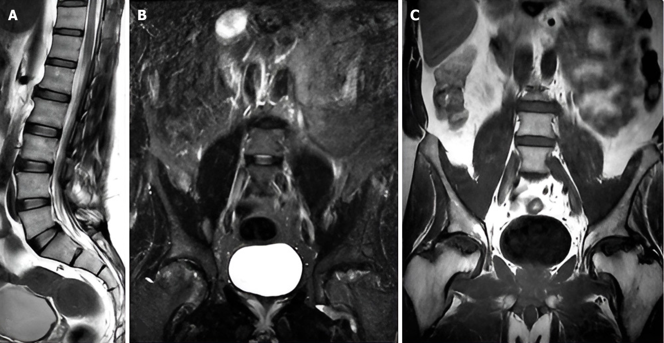
Figure 1 A 34-year-old male patient with low back and groin region pain.
A: Sagittal T2-weighted imaging showing muscular spasm; B and C: Coronal short tau inversion recovery and weighted imaging showing bone marrow edema of the femoral head bilaterally suggestive of Legg-Calve-Perthes disease.
- Citation: Al Kiswani S, Nasser M, Alzibdeh A, Lahham EE. Enhancing back pain and sciatica diagnosis: Coronal short tau inversion recovery’s role in routine lumbar magnetic resonance imaging protocols. World J Radiol 2025; 17(6): 107164
- URL: https://www.wjgnet.com/1949-8470/full/v17/i6/107164.htm
- DOI: https://dx.doi.org/10.4329/wjr.v17.i6.107164


