Copyright
©The Author(s) 2025.
World J Radiol. May 28, 2025; 17(5): 106975
Published online May 28, 2025. doi: 10.4329/wjr.v17.i5.106975
Published online May 28, 2025. doi: 10.4329/wjr.v17.i5.106975
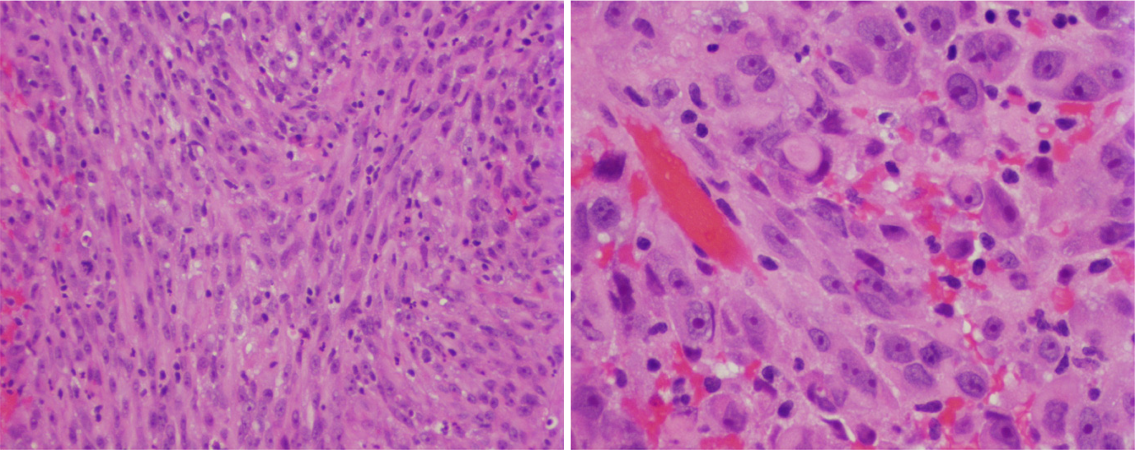
Figure 1 Histological features of sellar atypical teratoid/rhabdoid tumor.
Hematoxylin and eosin stain (Left). Epithelioid-like spindle cells with pink cytoplasm and prominent nuclei arranged in fascicular pattern, × 20 (Right). Focal tumor cells exhibiting rhabdoid features, × 40. Reproduced from Lev et al[16]. Citation: Lev I, Fan X, Yu R. Sellar Atypical Teratoid/Rhabdoid Tumor: Any Preoperative Diagnostic Clues? AACE Clin Case Rep 2015; 1: e2-e7. Copyright© 2015 Elsevier Inc. Published by Elsevier Inc. This is an open access article with User License: Creative Commons Attribution-NonCommercial-NoDerivs (CC BY-NC-ND 4.0). See: https://creativecommons.org/licenses/by-nc-nd/4.0/.
- Citation: Yu R. Specific imaging features of sellar atypical teratoid/rhabdoid tumor or the lack of thereof. World J Radiol 2025; 17(5): 106975
- URL: https://www.wjgnet.com/1949-8470/full/v17/i5/106975.htm
- DOI: https://dx.doi.org/10.4329/wjr.v17.i5.106975


