Copyright
©The Author(s) 2025.
World J Radiol. May 28, 2025; 17(5): 106102
Published online May 28, 2025. doi: 10.4329/wjr.v17.i5.106102
Published online May 28, 2025. doi: 10.4329/wjr.v17.i5.106102
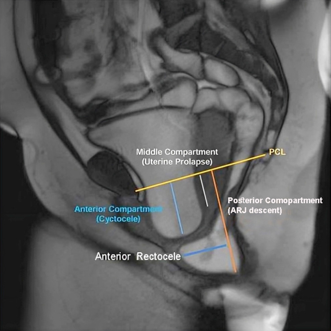
Figure 2 Magnetic resonance defecography image showing pelvic organ prolapse across all three pelvic compartments[15].
Anterior compartment: Cystocele (blue); Middle compartment: Uterine prolapse (white); Posterior compartment (orange, anterior rectocele and anorectal junction descent). Citation: Shetty A, Walizai T, Murphy A. MR defaecating proctography. Radiopaedia 2016. Copyright ©The Author(s) 2016. Published by Radiopaedia[15] (Supplementary material).
- Citation: Or-Rashid MH, Sultana A, Khanduker N, Ony TA, Hossain MM, Rahman J, Chowdhury MZ, Ahmed WU, Uddin MN, Uzzaman MS. Magnetic resonance defecography assessment of obstructed defecation syndrome in patients with chronic constipation in a tertiary care hospital. World J Radiol 2025; 17(5): 106102
- URL: https://www.wjgnet.com/1949-8470/full/v17/i5/106102.htm
- DOI: https://dx.doi.org/10.4329/wjr.v17.i5.106102


