
















©The Author(s) 2025.
World J Radiol. Nov 28, 2025; 17(11): 114754
Published online Nov 28, 2025. doi: 10.4329/wjr.v17.i11.114754
Published online Nov 28, 2025. doi: 10.4329/wjr.v17.i11.114754
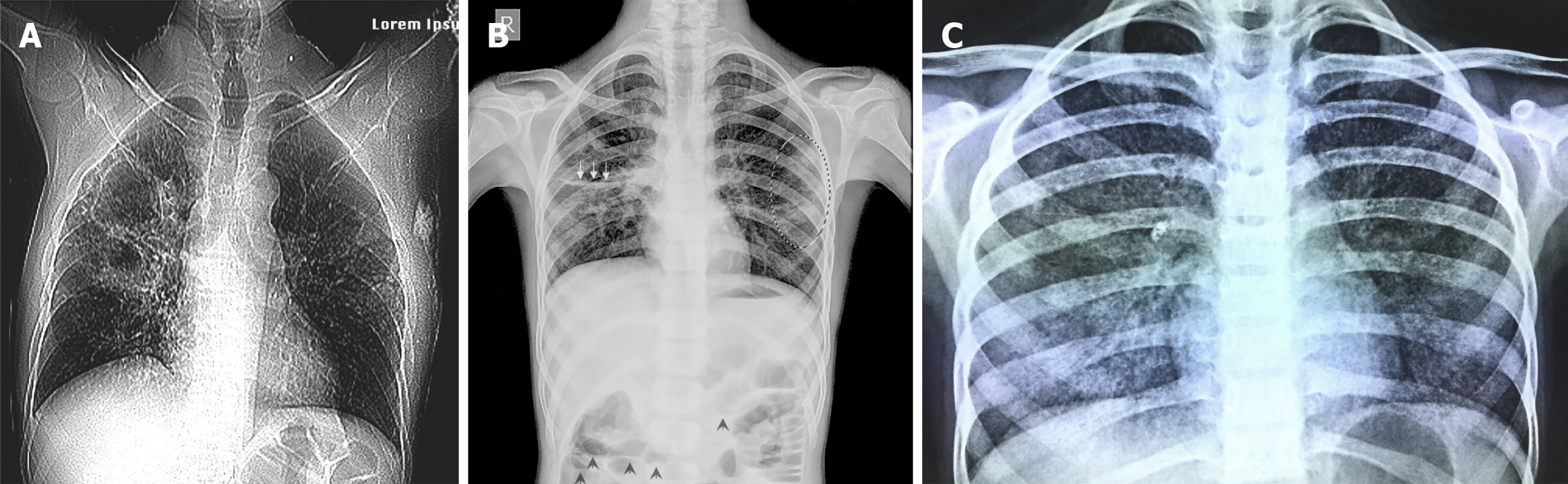
Figure 9 Frontal chest radiograph.
A: Frontal chest radiograph of a 14-year-old male presenting with cough and fever. Radiography demonstrates a thin-walled tuberculous cavity in the right upper lobe (white open arrowheads); B: Frontal chest radiograph of 9-year-old male with vomiting and abdominal pain, subsequently diagnosed with MDR tuberculosis (TB). Miliary pattern seen throughout both lung fields along with multiple larger lesions indicating multiple developing cavities (circled area), minimal effusion in the right horizontal fissure (white arrows) and hepatomegaly. Multiple air-fluid levels are seen in the visualized abdomen, signifying intestinal obstruction due to TB adhesions (short black arrows). (Figure 9A and B courtesy Dr. Jairaj Nair. Prof and HOD, Chest Medicine, LTMMC and LTMGH, Mumbai 400022); C: Frontal chest radiograph of 12-year-old boy with secondary hemophagocytic lymphohistiocytosis as a result of disseminated TB. Numerous miliary nodules are noted in both lungs (extensively) with additional patchy airspace opacities noted in the medial aspect of right lower lobe (note air-bronchogram). Calcified right hilar lymph nodes are also noted. He recovered well post treatment for same (including anti-TB treatment). (Image courtesy Dr. Prakash V Vaidya, Child Health Clinic, Mumbai and Sr. Consultant Pediatrician, Fortis Hospital, Mumbai 400080).
- Citation: Merchant SA, Merchant N, Varghese SL, Shaikh MJS. Large language models and large concept models in radiology: Present challenges, future directions, and critical perspectives. World J Radiol 2025; 17(11): 114754
- URL: https://www.wjgnet.com/1949-8470/full/v17/i11/114754.htm
- DOI: https://dx.doi.org/10.4329/wjr.v17.i11.114754