Copyright
©The Author(s) 2023.
World J Radiol. Mar 28, 2023; 15(3): 56-68
Published online Mar 28, 2023. doi: 10.4329/wjr.v15.i3.56
Published online Mar 28, 2023. doi: 10.4329/wjr.v15.i3.56
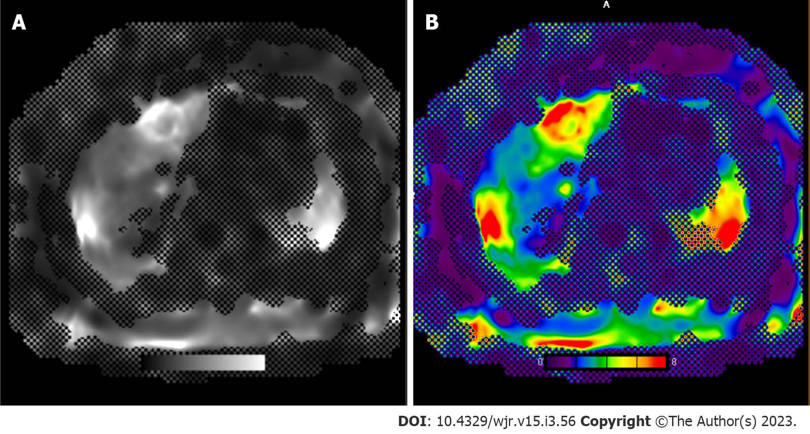
Figure 5 Magnetic resonance imaging elastography of the liver.
Resoundant driver system was used to induce acoustic vibrations in the liver, which were then tracked using magnetic resonance imaging (MRI) scanner to estimate hepatic stiffness. Gray scale and color-scale MRI elastography stiffness sequence. The mean liver stiffness is 4.0 kPa (range: 3.8-4.2 kPa). < 2.5 kPa = Normal; 0.5 to 2.93 kPa = Normal or inflammation; 2.93-3.5 kPa = Stage 1-2 fibrosis; 3.5-4 kPa = Stage 2-3 fibrosis; 4-5 kPa = Stage 3-4 fibrosis; > 5 kPa = Stage 4 or cirrhosis. A: Gray scale; B: Color-scale.
- Citation: Criss C, Nagar AM, Makary MS. Hepatocellular carcinoma: State of the art diagnostic imaging. World J Radiol 2023; 15(3): 56-68
- URL: https://www.wjgnet.com/1949-8470/full/v15/i3/56.htm
- DOI: https://dx.doi.org/10.4329/wjr.v15.i3.56


