
















©The Author(s) 2021.
World J Radiol. Jan 28, 2021; 13(1): 29-39
Published online Jan 28, 2021. doi: 10.4329/wjr.v13.i1.29
Published online Jan 28, 2021. doi: 10.4329/wjr.v13.i1.29
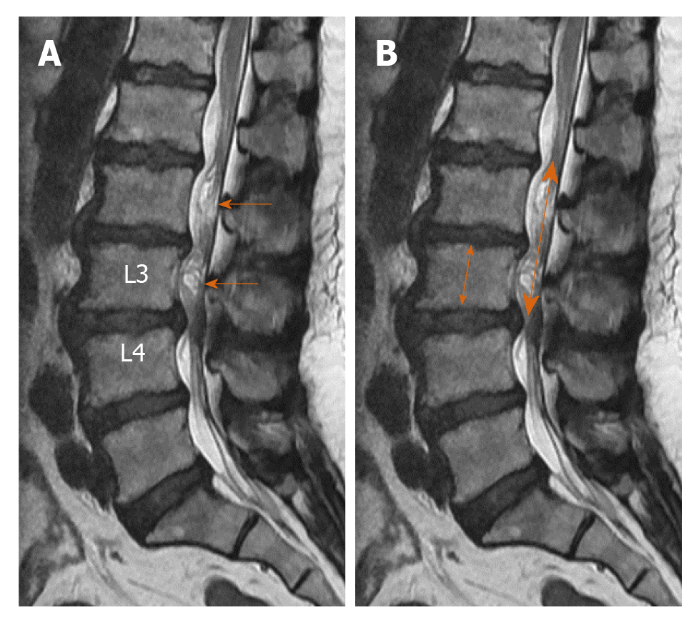
Figure 3 Forty-seven-year-old female patient with lumbar spondylosis.
A: On the sagittal T2-weighted image, redundant nerve roots at the superior of the stenosis level secondary to the stenosis at the L3-L4 intervertebral disc space (arrows) are shown; B: Relative length was calculated by dividing the length of redundant nerve roots (thick arrow) by the vertebra height at the superior of stenosis level (thin arrow).
- Citation: Gökçe E, Beyhan M. Magnetic resonance imaging findings of redundant nerve roots of the cauda equina. World J Radiol 2021; 13(1): 29-39
- URL: https://www.wjgnet.com/1949-8470/full/v13/i1/29.htm
- DOI: https://dx.doi.org/10.4329/wjr.v13.i1.29