Copyright
©The Author(s) 2020.
World J Radiol. Aug 28, 2020; 12(8): 156-171
Published online Aug 28, 2020. doi: 10.4329/wjr.v12.i8.156
Published online Aug 28, 2020. doi: 10.4329/wjr.v12.i8.156
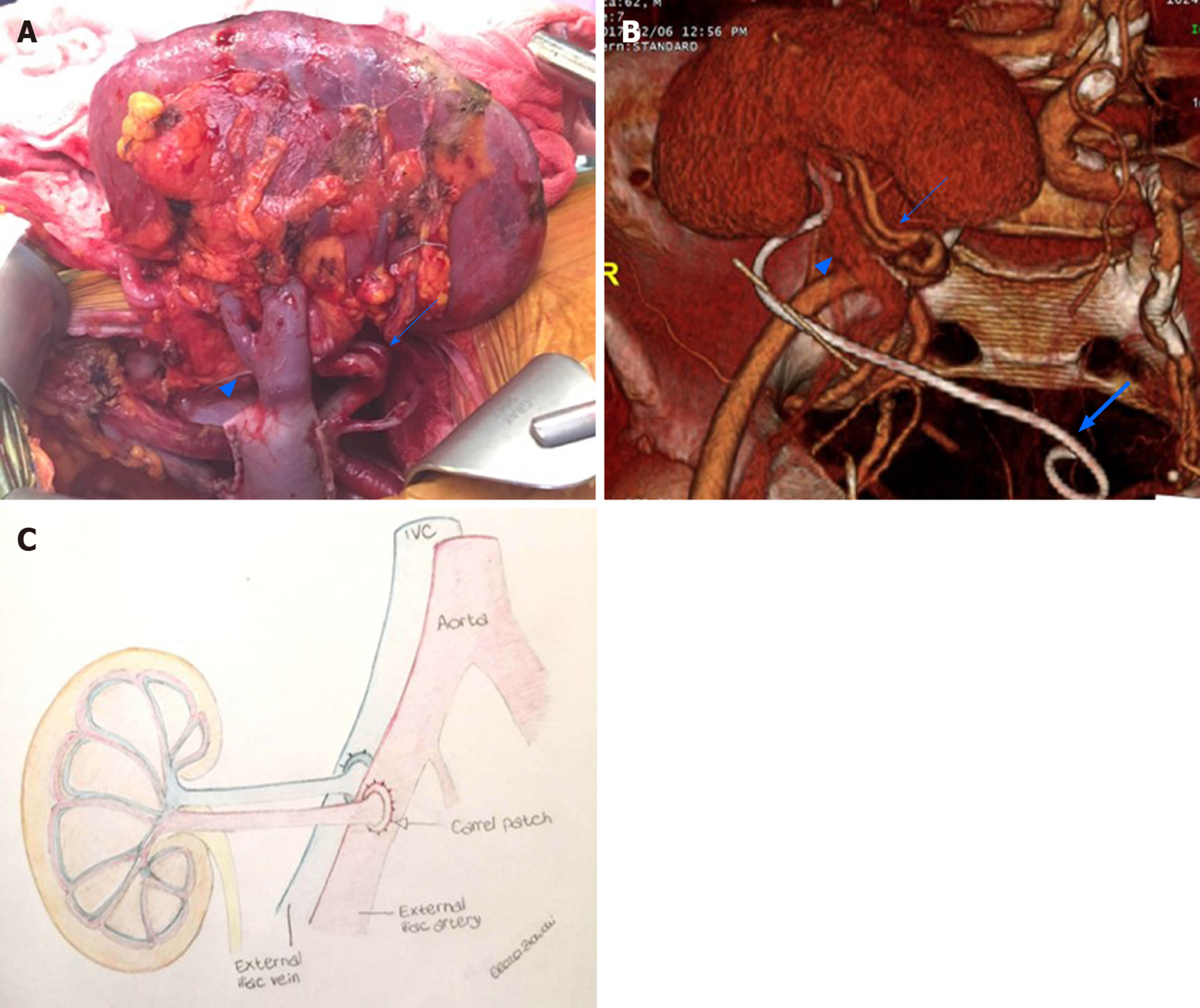
Figure 1 Renal allograft anatomy.
A: In the graft photo there is clear visualization of the transplanted renal vein (arrowhead) and transplanted renal artery (arrow); B: The corresponding volume rendering reconstruction from a computed tomography post-transplant scan shows the graft position in the right iliac fossa, transplanted renal vein (arrowhead), transplanted renal artery (arrow), and an ureteral stent temporarily left in place to favor urine output (large arrow); C: The drawing illustrates the end-to-end arterial anastomosis performed with a carrel patch (donor’s aortic patch attached to the transplanted renal artery) (arrow).
- Citation: Como G, Da Re J, Adani GL, Zuiani C, Girometti R. Role for contrast-enhanced ultrasound in assessing complications after kidney transplant. World J Radiol 2020; 12(8): 156-171
- URL: https://www.wjgnet.com/1949-8470/full/v12/i8/156.htm
- DOI: https://dx.doi.org/10.4329/wjr.v12.i8.156


