Copyright
©The Author(s) 2020.
World J Radiology. Mar 28, 2020; 12(3): 18-28
Published online Mar 28, 2020. doi: 10.4329/wjr.v12.i3.18
Published online Mar 28, 2020. doi: 10.4329/wjr.v12.i3.18
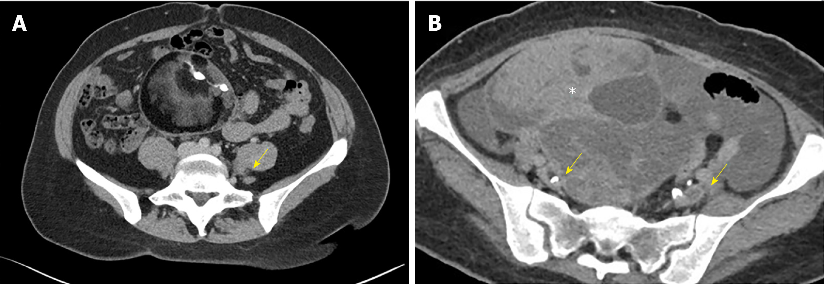
Figure 1 Axial Computed tomography venography image.
A: Axial computed tomography venography image showing compression of the left common iliac vein (arrow) between the right common iliac artery and the lumbar vertebral body; B: Extraluminal compression of the external iliac veins (arrows) from an ovarian tumor (asterixis).
- Citation: Toh MR, Tang TY, Lim HHMN, Venkatanarasimha N, Damodharan K. Review of imaging and endovascular intervention of iliocaval venous compression syndrome. World J Radiology 2020; 12(3): 18-28
- URL: https://www.wjgnet.com/1949-8470/full/v12/i3/18.htm
- DOI: https://dx.doi.org/10.4329/wjr.v12.i3.18


