
















©The Author(s) 2019.
World J Radiol. May 28, 2019; 11(5): 74-80
Published online May 28, 2019. doi: 10.4329/wjr.v11.i5.74
Published online May 28, 2019. doi: 10.4329/wjr.v11.i5.74
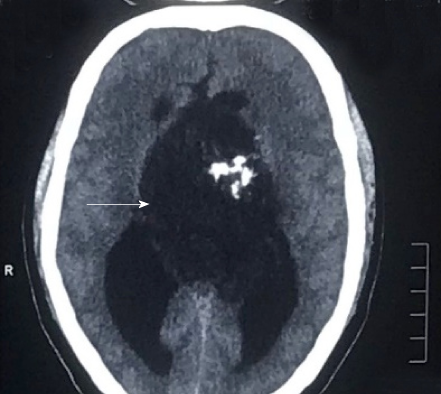
Figure 1 A non-contrast computed tomography scan of the head showing a mass arising from the third ventricle appearing hypodense and showing coarse nodular as well as curvilinear calcifications.
- Citation: Pawar S, Borde C, Patil A, Nagarkar R. Malignant epidermoid arising from the third ventricle: A case report. World J Radiol 2019; 11(5): 74-80
- URL: https://www.wjgnet.com/1949-8470/full/v11/i5/74.htm
- DOI: https://dx.doi.org/10.4329/wjr.v11.i5.74