Copyright
©The Author(s) 2019.
World J Radiol. Mar 28, 2019; 11(3): 27-45
Published online Mar 28, 2019. doi: 10.4329/wjr.v11.i3.27
Published online Mar 28, 2019. doi: 10.4329/wjr.v11.i3.27
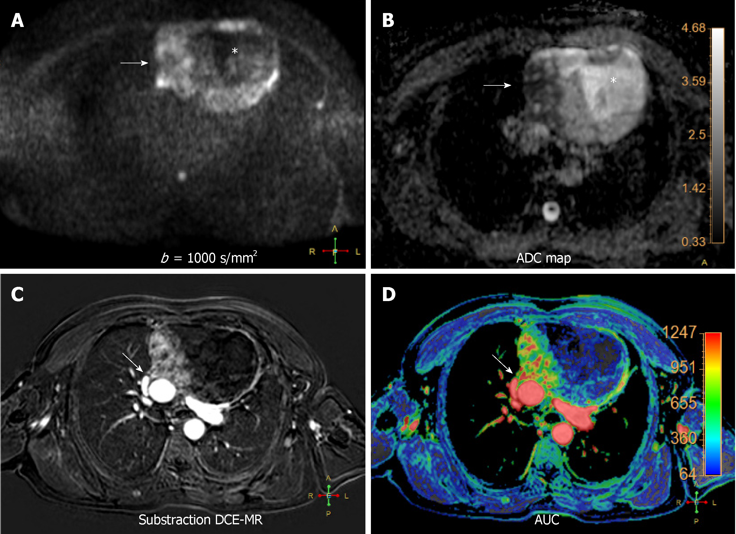
Figure 4 Multiparametric functional magnetic resonance imaging thymoepithelial mass.
A 63 year-old male with a complex cystic anterior mediastinal mass. A and B: High b value diffusion-weighted imaging (DWI) (b = 1000 s/mm2) (A) and corresponding apparent diffusion coefficient (ADC) map (B) revealing the complex behaviour of the lesion. In this case, DWI can differentiate the solid (white arrow on A and B) and cystic (white asterisk on A and B) components and also reveals heh restrictive behaviour of the solid part of the mass (ADC: 1.12-1.23 × 10⁻³ mm2/s), related to hypercellularity; C and D: Substraction of dynamic contrast-enhanced magnetic resonance on the arterial phase (C) and area under the curve parametric map (D) showing the locally invasive behaviour of the lesion to the pericardium and adjacent ascending aorta (white arrows on C and D). The mass corresponded to an invasive thymoma stage III of Masaoka–Koga classification system. DWI: Diffusion-weighted imaging; ADC: Apparent diffusion coefficient; AUC: Area under the curve; DCE-MR: Dynamic contrast-enhanced magnetic resonance.
- Citation: Broncano J, Alvarado-Benavides AM, Bhalla S, Álvarez-Kindelan A, Raptis CA, Luna A. Role of advanced magnetic resonance imaging in the assessment of malignancies of the mediastinum. World J Radiol 2019; 11(3): 27-45
- URL: https://www.wjgnet.com/1949-8470/full/v11/i3/27.htm
- DOI: https://dx.doi.org/10.4329/wjr.v11.i3.27


