
















©The Author(s) 2018.
World J Radiol. May 28, 2018; 10(5): 46-51
Published online May 28, 2018. doi: 10.4329/wjr.v10.i5.46
Published online May 28, 2018. doi: 10.4329/wjr.v10.i5.46
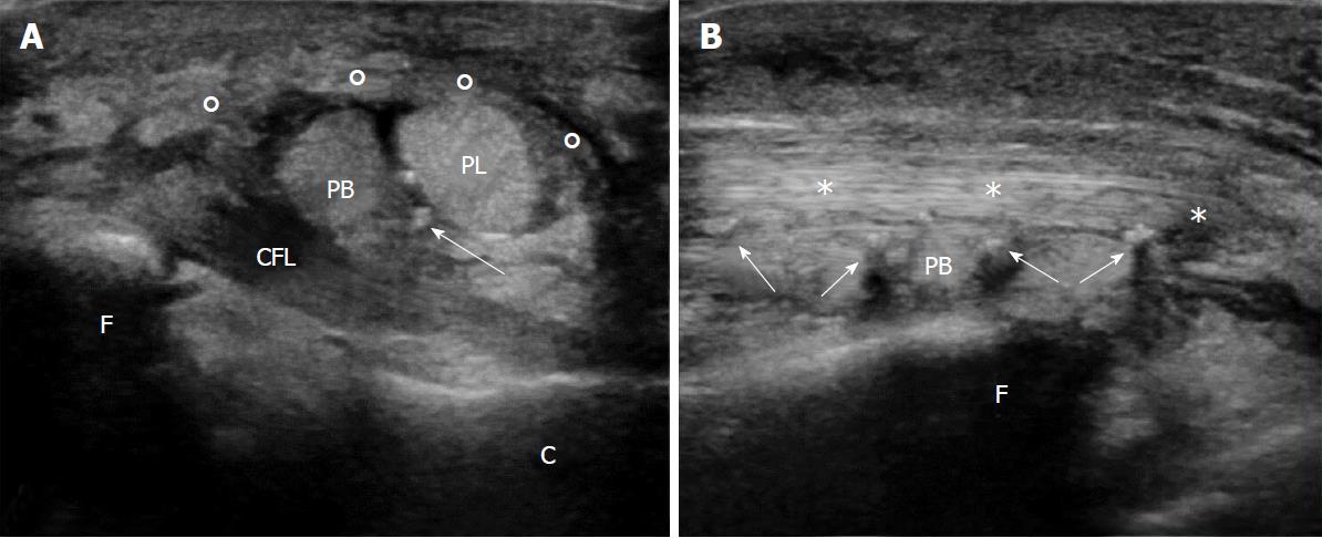
Figure 4 Post-surgical ultrasound examination of the injured ankle.
A: Axial US scan: the restored retinaculum (o) covers the two peroneus tendon, peroneus longus (PL) superiorly, and peroneus brevis (PB) inferiorly. Note the inhomogeneity of the lower part of the PB due to surgery. Suture stitches can be seen as hyperechoic spots (white arrows); fibula (F); calcaneus (C); calcaneofibular ligament (CFL); B: Longitudinal US scan. US: Ultrasound.
- Citation: Fischetti A, Zawaideh JP, Orlandi D, Belfiore S, SIlvestri E. Traumatic peroneal split lesion with retinaculum avulsion: Diagnosis and post-operative multymodality imaging. World J Radiol 2018; 10(5): 46-51
- URL: https://www.wjgnet.com/1949-8470/full/v10/i5/46.htm
- DOI: https://dx.doi.org/10.4329/wjr.v10.i5.46