Copyright
©2011 Baishideng Publishing Group Co.
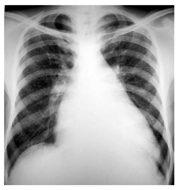
Figure 1 Chest radiography showed cardiomegaly with straightening of the left heart border.
In addition, there were diffusely scattered miliary nodular opacities (approximately 3 mm).
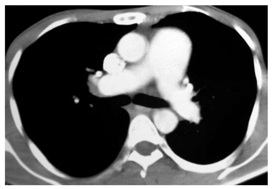
Figure 2 Chest computed tomography (mediastinal sections) showed an enlarged main pulmonary arterial trunk.
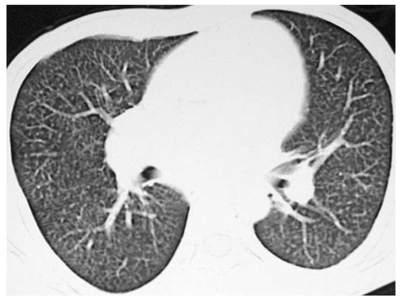
Figure 3 Chest computed tomography (lung windows) showed randomly scattered miliary nodules.
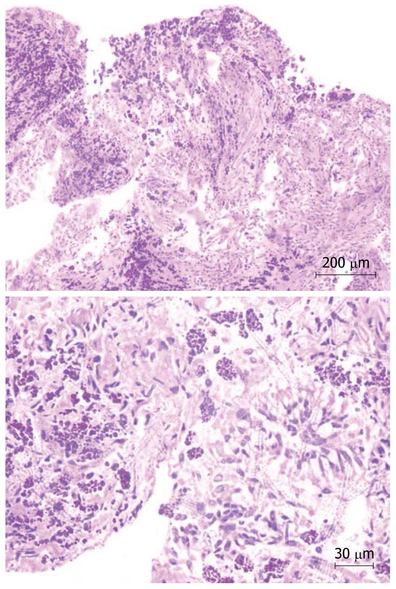
Figure 4 Bronchoscopic lung biopsy (upper) showed alveoli filled with coarse pigment-laden macrophages with sparse lymphocytic infiltrate in the interstitial septa.
The figure on the bottom reveals the same findings at a higher magnification.
- Citation: Agrawal G, Agarwal R, Rohit MK, Mahesh V, Vasishta RK. Miliary nodules due to secondary pulmonary hemosiderosis in rheumatic heart disease. World J Radiol 2011; 3(2): 51-54
- URL: https://www.wjgnet.com/1949-8470/full/v3/i2/51.htm
- DOI: https://dx.doi.org/10.4329/wjr.v3.i2.51


