Copyright
©The Author(s) 2022.
World J Radiol. Apr 28, 2022; 14(4): 91-103
Published online Apr 28, 2022. doi: 10.4329/wjr.v14.i4.91
Published online Apr 28, 2022. doi: 10.4329/wjr.v14.i4.91
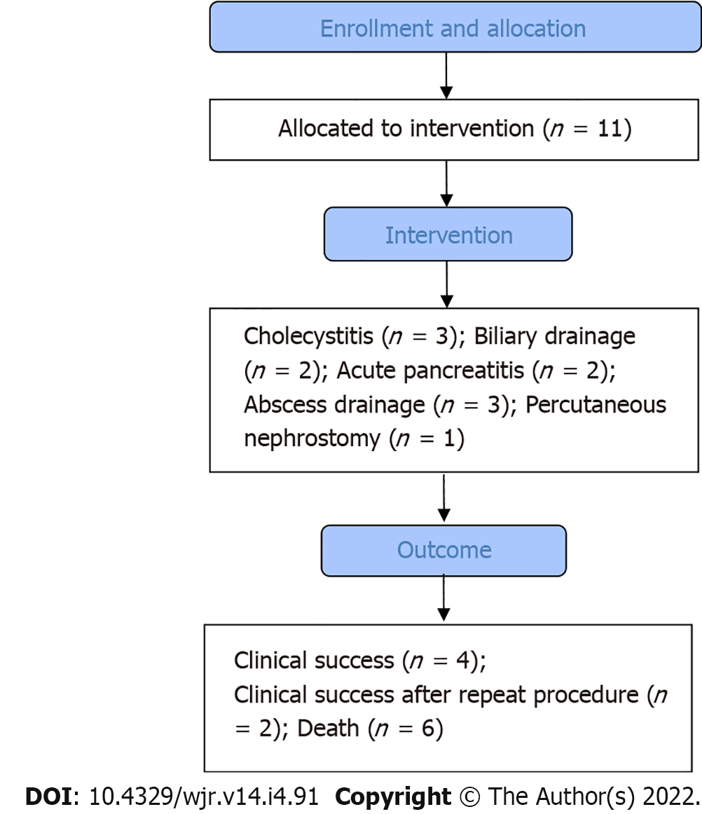
Figure 1 Flow chart of the study.
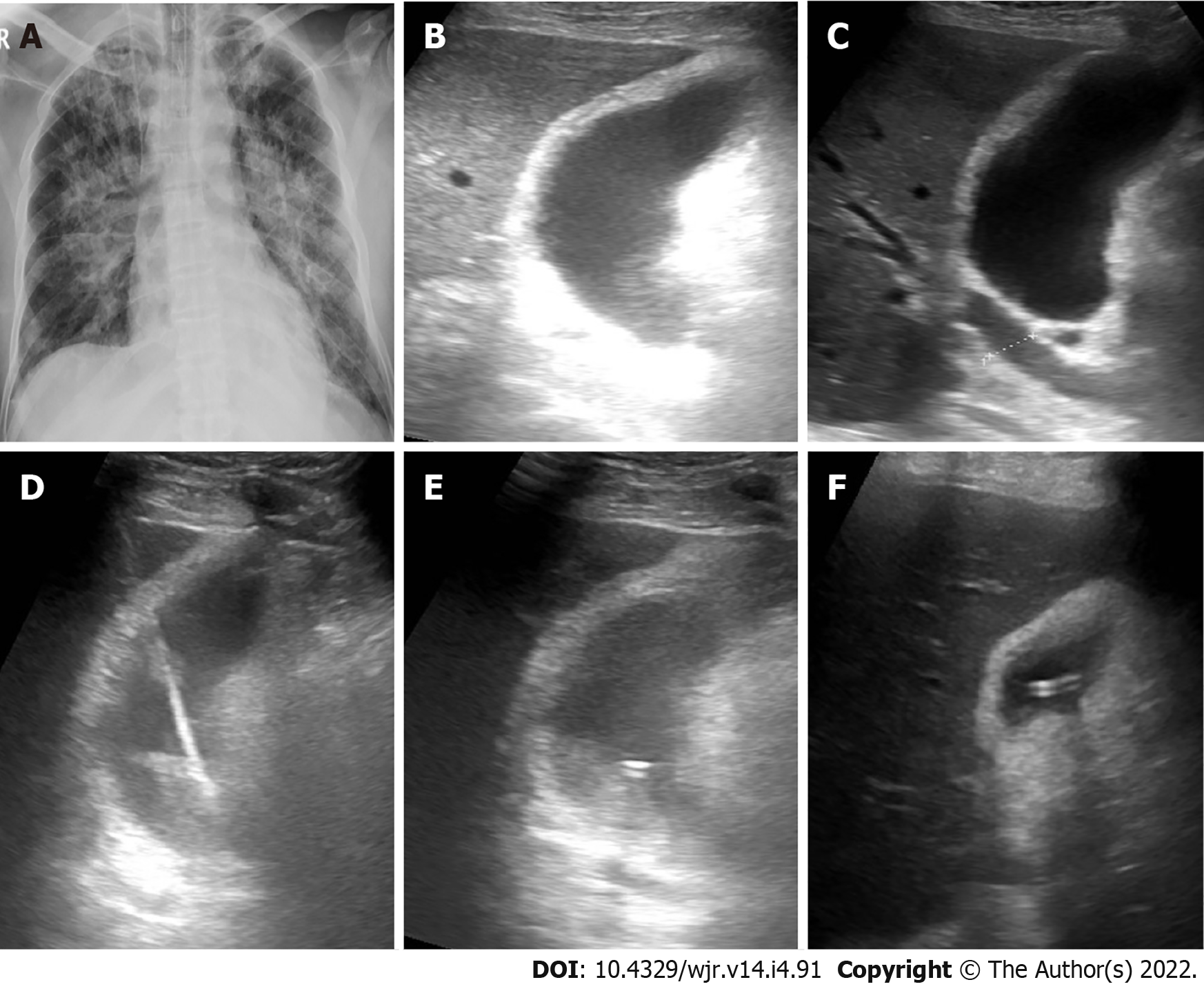
Figure 2 Cholecystostomy in a 72-yr-old male presented by acute cholecystitis.
A: Frontal chest X-ray shows opacities involving both lungs with central predominance; B and C: B-mode ultrasound images show distended thick-walled gall bladder with biliary dilatation; D: B-mode ultrasound image show puncture needle through the gall bladder; E: B-mode ultrasound image tube inside the gall bladder; F: B-mode ultrasound image of the gall bladder after drainage.
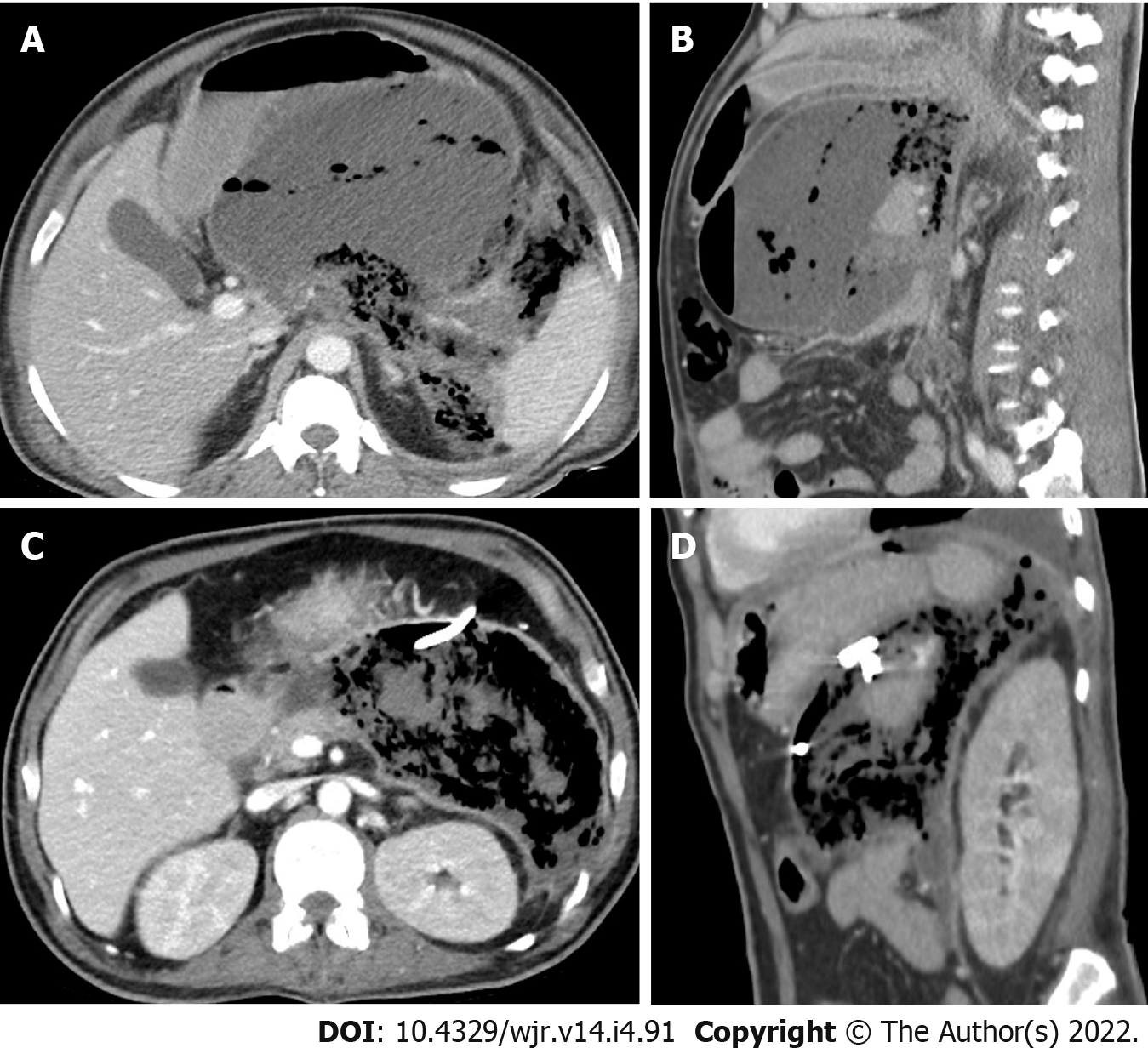
Figure 3 Percutaneous drainage of peripancreatic collection in a 43-yr-old male presented by acute pancreatitis.
A and B: Axial and sagittal contrast enhanced computed tomography (CT) images show large peripancreatic collection/walled-off necrosis. The collection is mixed with pockets of gas inside and there is extension of the gas density into the retroperitoneal and perisplenic spaces; C and D: Axial and sagittal contrast enhanced CT images 22 d after tube insertion show reduction of the collection size with increased amount of gas within the collection.
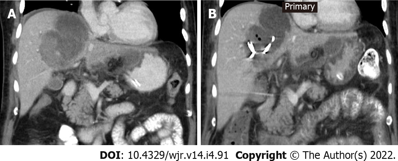
Figure 4 Percutaneous drainage of hepatic abscess in a 63-yr-old male.
A: Coronal contrast enhanced computed tomography (CT) image shows thick-walled hepatic abscess with dependent high density inside secondary to clotted blood, a rim of perihepatic fluid is also noted; B: Coronal contrast enhanced CT image 6 d after tube insertion show reduction of the abscess size with few foci of gas density.
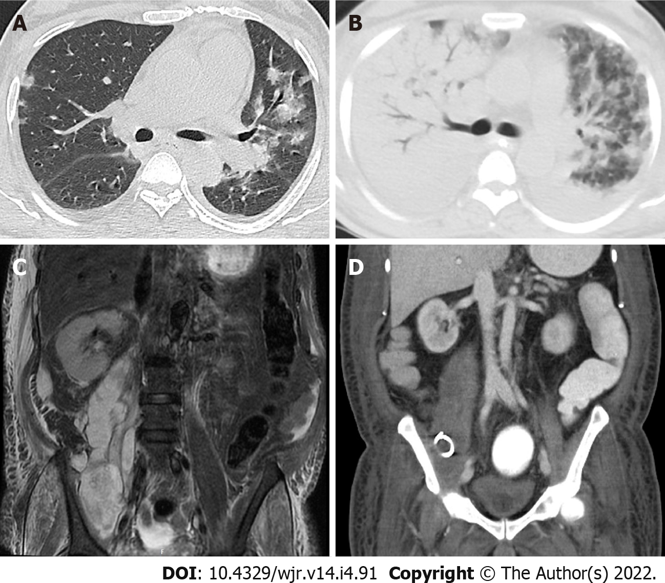
Figure 5 Percutaneous drainage of right psoas major abscess in a 60-yr-old male.
A: Axial chest computed tomography (CT) image in pulmonary window shows bilateral ground-glass opacities (GGOs) and minimal bilateral pleural effusion; B: Axial chest CT image in pulmonary window 11 d after initial CT shows bilateral consolidation involving most of the right lung and GGOs in the remaining left lung parenchyma; C: Coronal T2 FAT SAT image shows large multi-locular psoas major abscess associated with muscular and subcutaneous soft tissue edema; D: Coronal contrast enhanced CT images 8 d after tube insertion show reduction of the collection size with regression of the associated soft tissue edema.
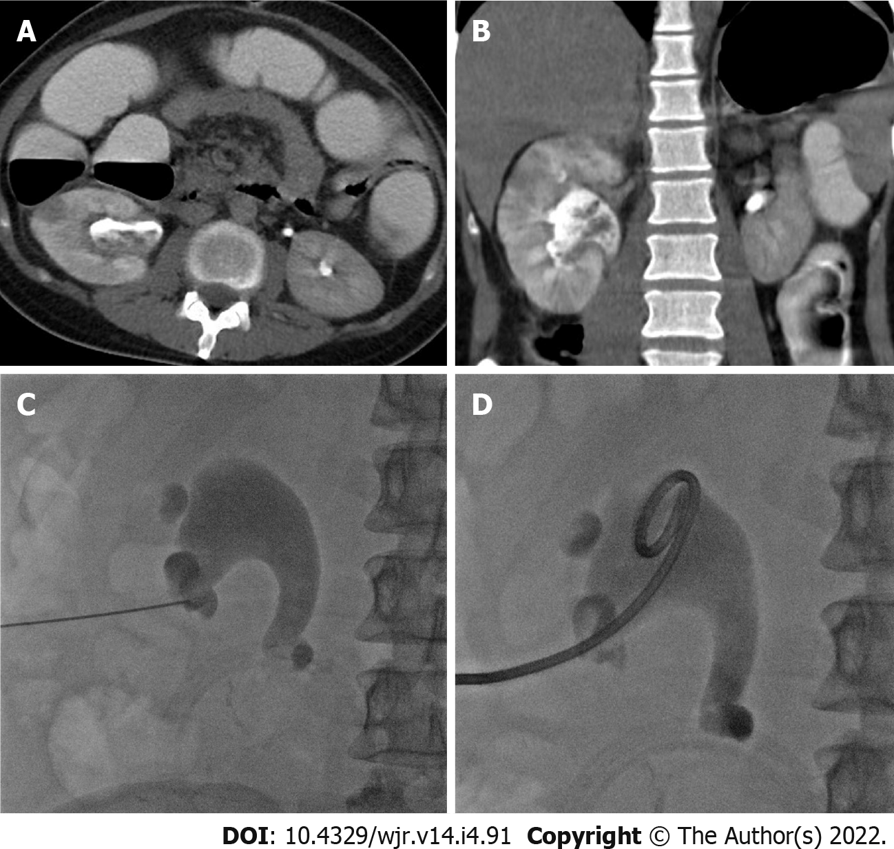
Figure 6 Percutaneous nephrostomy in a 30-yr-old male presented with acute pyelonephritis.
A and B: Axial and coronal computed tomography images in excretory phase show characteristic features of acute pyelonephritis in the form of focal hypoenhnacing areas (striated nephrogram) and debris in dilated renal pelvis; C and D: Frontal fluoroscopic images show puncture needle in the lower calyces and successful insertion of nephrostomy tube.
- Citation: Deif MA, Mounir AM, Abo-Hedibah SA, Abdel Khalek AM, Elmokadem AH. Outcome of percutaneous drainage for septic complications coexisted with COVID-19. World J Radiol 2022; 14(4): 91-103
- URL: https://www.wjgnet.com/1949-8470/full/v14/i4/91.htm
- DOI: https://dx.doi.org/10.4329/wjr.v14.i4.91


