Copyright
©The Author(s) 2017.
World J Cardiol. Feb 26, 2017; 9(2): 92-108
Published online Feb 26, 2017. doi: 10.4330/wjc.v9.i2.92
Published online Feb 26, 2017. doi: 10.4330/wjc.v9.i2.92
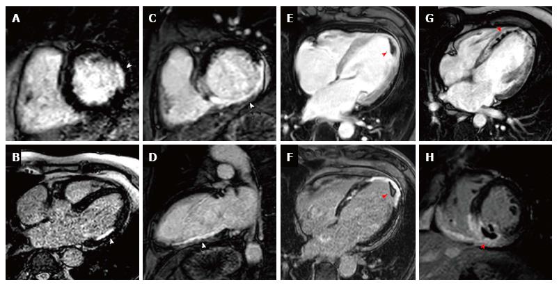
Figure 4 Early and late gadolinium enhancement.
A and B show a lateral sub-endocardial infarction on short axis and 4 chamber LGE respectively; C and D show a full thickness inferior infarction on LGE imaging on short axis and VLA respectively; E and F show EGE and LGE imaging respectively of a full thickness apical infarction with an apical thrombus appearing black (highlighted by red arrow); G shows an extensive acute antero-apical infarction with a core of microvascular obstruction visible within the hyperenhancement on EGE (red arrow); H shows an acute inferior wall infarction with MVO and extension into the right ventricle on LGE (red arrow) imaging. LGE: Late gadolinium enhancement; EGE: Early gadolinium enhancement; MVO: Mitral orifice.
- Citation: Foley JRJ, Plein S, Greenwood JP. Assessment of stable coronary artery disease by cardiovascular magnetic resonance imaging: Current and emerging techniques. World J Cardiol 2017; 9(2): 92-108
- URL: https://www.wjgnet.com/1949-8462/full/v9/i2/92.htm
- DOI: https://dx.doi.org/10.4330/wjc.v9.i2.92


