Copyright
©The Author(s) 2017.
World J Cardiol. Feb 26, 2017; 9(2): 92-108
Published online Feb 26, 2017. doi: 10.4330/wjc.v9.i2.92
Published online Feb 26, 2017. doi: 10.4330/wjc.v9.i2.92
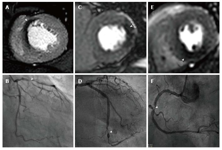
Figure 3 Cardiovascular magnetic resonance perfusion techniques.
A is a high spatial resolution k-t BLAST stress perfusion CMR study at 3.0T showing an antero-septal perfusion defect with corresponding left anterior descending lesion at angiography in B; C shows a transmural lateral perfusion defect at standard resolution at 1.5T with corresponding circumflex lesion in D; E shows a transmural inferior perfusion defect at standard resolution at 1.5T with corresponding right coronary artery lesion in F. BLAST: Broad-use linear acquisition speed-up technique; CMR: Cardiovascular magnetic resonance.
- Citation: Foley JRJ, Plein S, Greenwood JP. Assessment of stable coronary artery disease by cardiovascular magnetic resonance imaging: Current and emerging techniques. World J Cardiol 2017; 9(2): 92-108
- URL: https://www.wjgnet.com/1949-8462/full/v9/i2/92.htm
- DOI: https://dx.doi.org/10.4330/wjc.v9.i2.92


