
















©The Author(s) 2017.
World J Cardiol. Dec 26, 2017; 9(12): 853-857
Published online Dec 26, 2017. doi: 10.4330/wjc.v9.i12.853
Published online Dec 26, 2017. doi: 10.4330/wjc.v9.i12.853
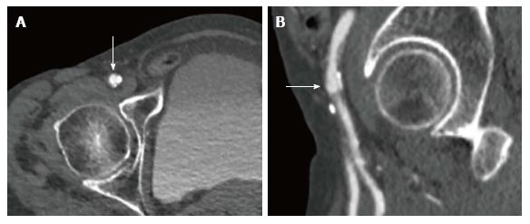
Figure 4 Multislice computed tomography showing calcified right common femoral artery in a patient undergoing transfemoral transcatheter aortic valve implantation.
A: Right common femoral artery with an arrow pointing at the ideal puncture site above the calcification; B: Right common femoral artery with an arrow pointing at the ideal puncture site above the height of bifurcation of the common femoral artery in relationship to the femoral head.
- Citation: Brinkert M, Toggweiler S. Transcatheter aortic valve implantation operators - get involved in imaging! World J Cardiol 2017; 9(12): 853-857
- URL: https://www.wjgnet.com/1949-8462/full/v9/i12/853.htm
- DOI: https://dx.doi.org/10.4330/wjc.v9.i12.853