Copyright
©The Author(s) 2017.
World J Cardiol. Dec 26, 2017; 9(12): 853-857
Published online Dec 26, 2017. doi: 10.4330/wjc.v9.i12.853
Published online Dec 26, 2017. doi: 10.4330/wjc.v9.i12.853
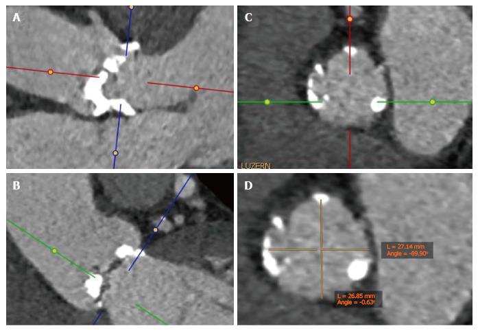
Figure 1 Example of a multiplanar reconstruction of the aortic annulus.
A and B: Double-oblique MSCT images at the basal insertion of the calcified native cusps; C: Double-oblique reconstruction at the level of the aortic annulus. The aortic valve leaflets are just barely visible at the level of the ventriculoarterial junction; D: Measurement of the short and long diameter at the level of the aortic annulus. MSCT: Multislice computed tomography.
- Citation: Brinkert M, Toggweiler S. Transcatheter aortic valve implantation operators - get involved in imaging! World J Cardiol 2017; 9(12): 853-857
- URL: https://www.wjgnet.com/1949-8462/full/v9/i12/853.htm
- DOI: https://dx.doi.org/10.4330/wjc.v9.i12.853


