Copyright
©The Author(s) 2016.
World J Cardiol. Nov 26, 2016; 8(11): 623-637
Published online Nov 26, 2016. doi: 10.4330/wjc.v8.i11.623
Published online Nov 26, 2016. doi: 10.4330/wjc.v8.i11.623
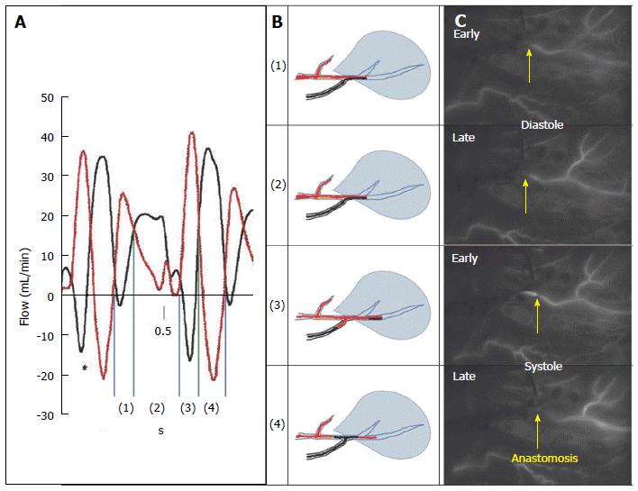
Figure 5 Intraoperative real-time documentation of competitive flow in arterial internal mammary artery-left anterior descending coronary anastomosis with > 70% stenosis.
Competitive flow documented in in situ arterial graft to TVECA with > 70% proximal stenosis. A: Dynamic flow data from Pagni et al[71] illustrating flow in IMA (red) and LAD (black) in an experimental model of competitive flow where the LAD has no proximal stenosis (maximal competitive flow). In phases 1 and 2, during diastole, there is antegrade flow in both the TVECA and the arterial graft. In early systole, there is antegrade flow in the TVECA but retrograde flow in the distal arterial graft, which reverses in late systole, where there is retrograde flow in the TVECA and antegrade flow in the arterial conduit; B: Diagrammatically the IMA-LAD interaction at the anastomosis in this patient; C: Four still frames taken from the 34-s video of this bypass graft using near-infrared imaging technology (SPY, Novadaq Technologies, Toronto, Ontario, CA). The arrow indicates the site of the anastomosis. The four frames are in temporal sequence but not consecutive frames; they are selected at the four diagram points indicated at the middle panel. Each diagram point is taken from the appropriate time-point within each of the four intervals (early diastole, late diastole, early systole, late diastole). This real-time intraoperative imaging shows identical flow patterns as demonstrated in the Pagni experimental model, despite the proximal > 70% stenosis. IMA: Internal mammary artery; LAD: Left anterior descending coronary artery; TVECA: Target vessel epicardial coronary artery.
- Citation: Ferguson Jr TB. Physiology of in-situ arterial revascularization in coronary artery bypass grafting: Preoperative, intraoperative and postoperative factors and influences. World J Cardiol 2016; 8(11): 623-637
- URL: https://www.wjgnet.com/1949-8462/full/v8/i11/623.htm
- DOI: https://dx.doi.org/10.4330/wjc.v8.i11.623


