Copyright
©2014 Baishideng Publishing Group Inc.
World J Cardiol. Sep 26, 2014; 6(9): 959-967
Published online Sep 26, 2014. doi: 10.4330/wjc.v6.i9.959
Published online Sep 26, 2014. doi: 10.4330/wjc.v6.i9.959
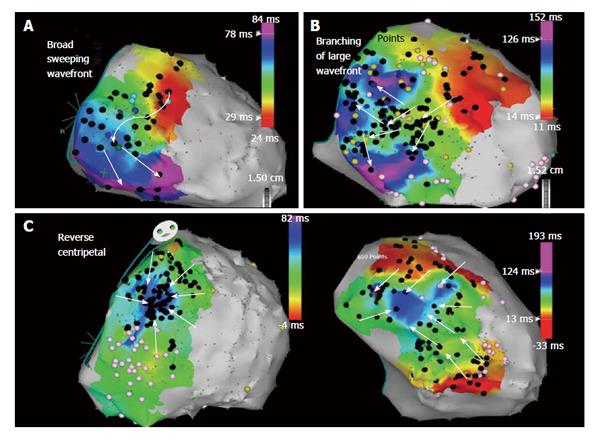
Figure 7 Epicardial right ventricle free wall activation maps illustrating propagation wavefront of epicardial isolated potentials in a patient with arrhythmogenic right ventricular cardiomyopathy/dysplasia.
A: Right ventricle (RV) free wall activation via a broad wavefront progressing toward the inferior RV; B: A diverging pattern of activation, initially broad, but subsequently branching as it progresses through the scar; C: Reverse centripetal pattern with outside activation progressing inward with wavefront collision in the center of the scar. Adapted from Haqqani et al[18] with permission.
- Citation: Tschabrunn CM, Marchlinski FE. Ventricular tachycardia mapping and ablation in arrhythmogenic right ventricular cardiomyopathy/dysplasia: Lessons Learned. World J Cardiol 2014; 6(9): 959-967
- URL: https://www.wjgnet.com/1949-8462/full/v6/i9/959.htm
- DOI: https://dx.doi.org/10.4330/wjc.v6.i9.959


