Copyright
©2014 Baishideng Publishing Group Inc.
World J Cardiol. Sep 26, 2014; 6(9): 959-967
Published online Sep 26, 2014. doi: 10.4330/wjc.v6.i9.959
Published online Sep 26, 2014. doi: 10.4330/wjc.v6.i9.959
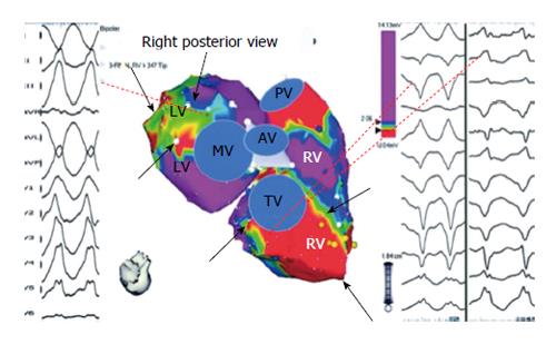
Figure 2 Bipolar right ventricle and left ventricle endocardial voltage maps highlighting location of abnormal endocardium and origin of ventricular tachycardia in a patient with right ventricle cardiomyopathy and ventricular tachycardia.
Electroanatomic abnormalities include both the tricuspid and mitral valves from tricuspid and mitral valves (black arrows). Origin of ventricular tachycardia (VT) based on activation and pace mapping was perivalvular mitral for the RBBB VT and perivalvular tricuspid valve for LBBB VT (dashed lines). Adapted from Marchlinski et al[15] with permission. RV: Right ventricle; LV: Left ventricle.
- Citation: Tschabrunn CM, Marchlinski FE. Ventricular tachycardia mapping and ablation in arrhythmogenic right ventricular cardiomyopathy/dysplasia: Lessons Learned. World J Cardiol 2014; 6(9): 959-967
- URL: https://www.wjgnet.com/1949-8462/full/v6/i9/959.htm
- DOI: https://dx.doi.org/10.4330/wjc.v6.i9.959


