Copyright
©2014 Baishideng Publishing Group Inc.
World J Cardiol. Aug 26, 2014; 6(8): 771-781
Published online Aug 26, 2014. doi: 10.4330/wjc.v6.i8.771
Published online Aug 26, 2014. doi: 10.4330/wjc.v6.i8.771
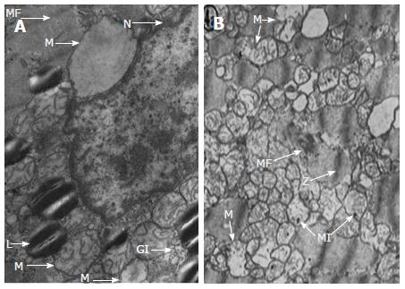
Figure 2 Cellular changes in alcoholic cardiomyopathy.
L: Neutral lipids in the form of small cytoplasmic droplets; GI: Glycogen deposits; M: Mitochondria were swollen or oedema was present; N: Nucleus; MF: Myofibrils showed a progressively distorted structure (Z lines disrupted). Reproduced with permission from the American Heart Association[42].
- Citation: Guzzo-Merello G, Cobo-Marcos M, Gallego-Delgado M, Garcia-Pavia P. Alcoholic cardiomyopathy. World J Cardiol 2014; 6(8): 771-781
- URL: https://www.wjgnet.com/1949-8462/full/v6/i8/771.htm
- DOI: https://dx.doi.org/10.4330/wjc.v6.i8.771


