
















©2013 Baishideng Publishing Group Co.
World J Cardiol. Apr 26, 2013; 5(4): 109-111
Published online Apr 26, 2013. doi: 10.4330/wjc.v5.i4.109
Published online Apr 26, 2013. doi: 10.4330/wjc.v5.i4.109
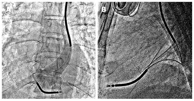
Figure 2 X-ray in Antero-posterior (A) and lateral (B) view shows dual coil right ventricle lead implanted at right ventricle apex.
Venogram in figure (A) confirms persistence of left superior vena cava.
- Citation: Vijayvergiya R, Shrivastava S, Kumar A, Otaal PS. Transvenous defibrillator implantation in a patient with persistent left superior vena cava. World J Cardiol 2013; 5(4): 109-111
- URL: https://www.wjgnet.com/1949-8462/full/v5/i4/109.htm
- DOI: https://dx.doi.org/10.4330/wjc.v5.i4.109