Copyright
©2013 Baishideng Publishing Group Co.
World J Cardiol. Dec 26, 2013; 5(12): 473-483
Published online Dec 26, 2013. doi: 10.4330/wjc.v5.i12.473
Published online Dec 26, 2013. doi: 10.4330/wjc.v5.i12.473
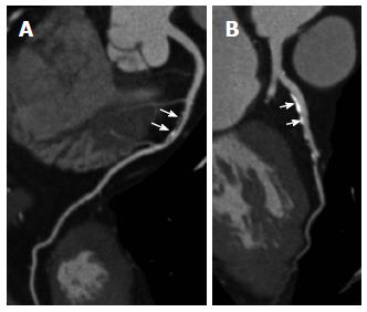
Figure 3 Electrocardiogram-triggered coronary computed tomography angiography.
Prospectively ECG-triggered coronary computed tomography angiography shows a mixed plaque at the mid-segment of right coronary artery (A, arrows), and calcified plaques at the proximal segment of left anterior descending branch (B, arrows). ECG: Electrocardiogram.
- Citation: Sabarudin A, Sun Z. Coronary CT angiography: Diagnostic value and clinical challenges. World J Cardiol 2013; 5(12): 473-483
- URL: https://www.wjgnet.com/1949-8462/full/v5/i12/473.htm
- DOI: https://dx.doi.org/10.4330/wjc.v5.i12.473


