Copyright
©2010 Baishideng Publishing Group Co.
World J Cardiol. Jul 26, 2010; 2(7): 163-170
Published online Jul 26, 2010. doi: 10.4330/wjc.v2.i7.163
Published online Jul 26, 2010. doi: 10.4330/wjc.v2.i7.163
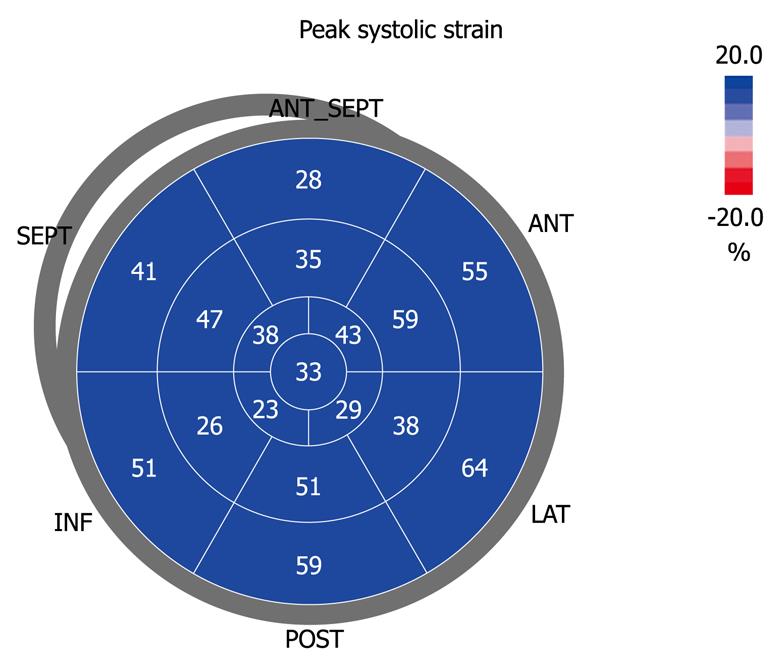
Figure 5 Bull’s eye view of the 2D strain of the left atrial reservoir in a normal subject.
Bull’s eye with the longitudinal 2D strain values of the left atrium. It is colored blue because it represents the atrial strain of the reservoir phase, which increases during ventricular systole. Note that basal values are higher than medial values and further reduced at the center, which represents the atrial roof strain. Values of the antero-septal wall should be excluded, because they correspond to the ascending aorta.
- Citation: Cianciulli TF, Saccheri MC, Lax JA, Bermann AM, Ferreiro DE. Two-dimensional speckle tracking echocardiography for the assessment of atrial function. World J Cardiol 2010; 2(7): 163-170
- URL: https://www.wjgnet.com/1949-8462/full/v2/i7/163.htm
- DOI: https://dx.doi.org/10.4330/wjc.v2.i7.163


