
















Copyright: ©Author(s) 2026.
World J Cardiol. Apr 26, 2026; 18(4): 118863
Published online Apr 26, 2026. doi: 10.4330/wjc.v18.i4.118863
Published online Apr 26, 2026. doi: 10.4330/wjc.v18.i4.118863
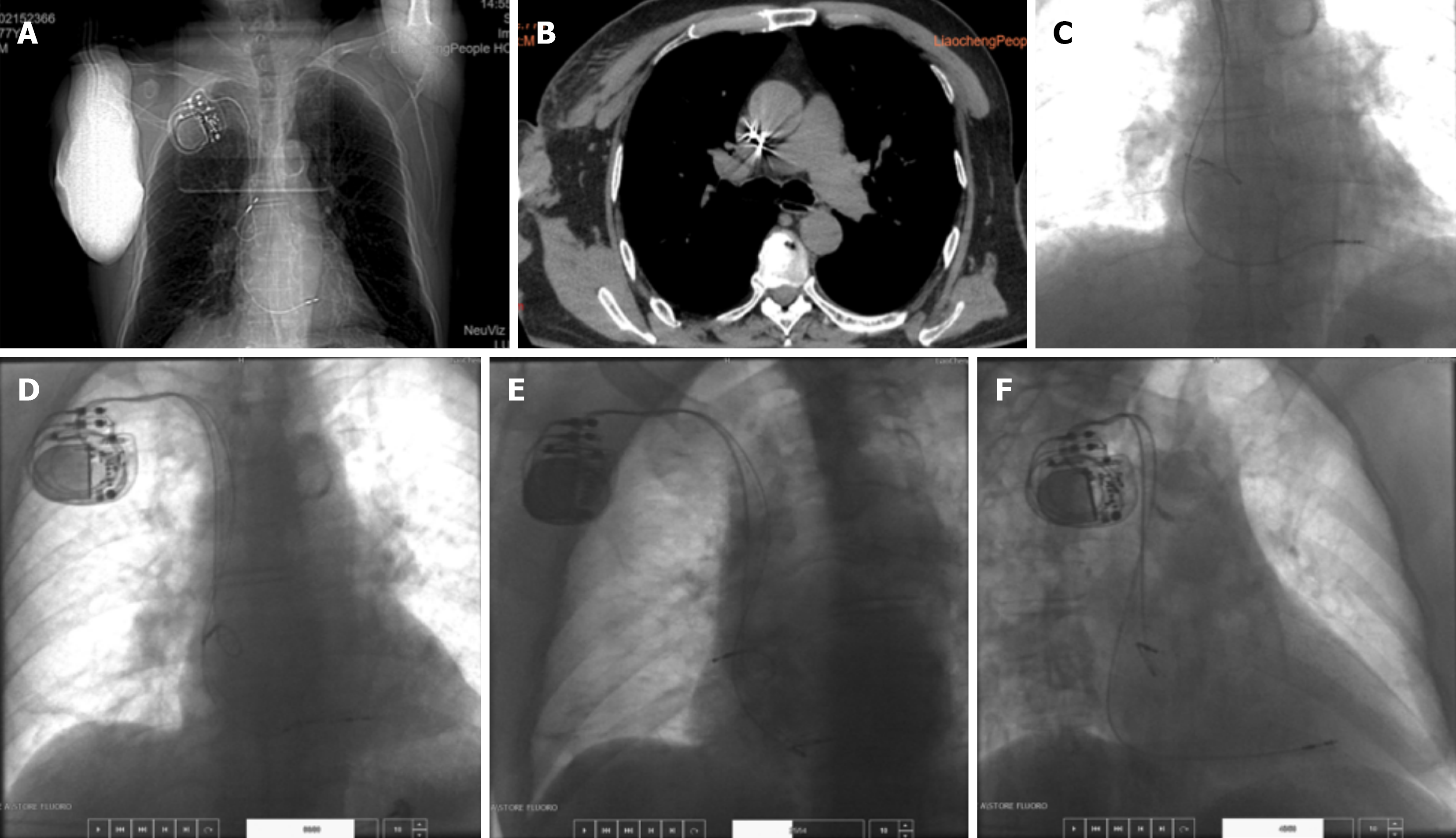
Figure 2 Changes in the position of the pacemaker lead.
A: Anteroposterior chest X-ray showing no obvious change in the ventricular lead compared with the previous image, while the atrial lead is directed leftward; B: Computed tomography scan. The arrow indicates the tip of the dislodged atrial lead, pointing toward the junction of the posterior atrial wall and the superior vena cava; C: Anteroposterior X-ray image after electrode placement and before lead fixation. The ventricular active lead is fixed at the mid-septum. The helix was unscrewed 13 turns, and the screw was fully unscrewed. The ventricular lead was well secured with appropriate slack. The atrial passive lead points toward the right atrial appendage; D: Anteroposterior view showing no obvious change in the position of the ventricular lead compared with Figure 2C. The atrial lead has rotated clockwise compared with Figure 2C, but the tip still points toward the right atrial appendage; E: Left anterior oblique view showing that the ventricular lead directed toward the septum, with the atrial lead tip pointing to the right atrial appendage; F: Right anterior oblique view showing that the atrial lead tip directed anteriorly and inferiorly.
- Citation: Zhang XS, Wang F, Qi XW, Shafii P, Cui XN, Wang YH. Phrenic nerve capture caused by atrial electrode dislodgement: A case report. World J Cardiol 2026; 18(4): 118863
- URL: https://www.wjgnet.com/1949-8462/full/v18/i4/118863.htm
- DOI: https://dx.doi.org/10.4330/wjc.v18.i4.118863