
















Copyright: ©Author(s) 2026.
World J Cardiol. Mar 26, 2026; 18(3): 117999
Published online Mar 26, 2026. doi: 10.4330/wjc.v18.i3.117999
Published online Mar 26, 2026. doi: 10.4330/wjc.v18.i3.117999
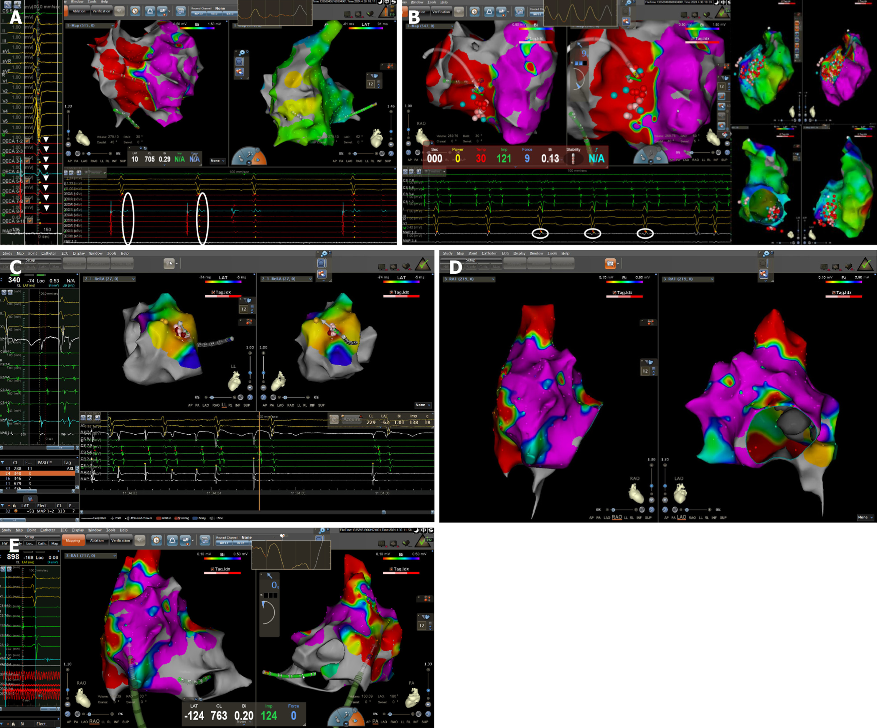
Figure 4 Electroanatomic mapping and ablation.
A: Low-frequency, fractionated late potentials; B: Homogenization modification; C: Para-Hisian focal atrial tachycardia ablation; D and E: Extensive low-voltage and scarred areas on the right atrial free wall, posterior wall, and the sinus-node region, corresponding to the signal of cardiac magnetic resonance in Figure 3B.
- Citation: Li A, Zhu YJ, Ma XX, Jin Y, Yang J, Feng QT. Arrhythmogenic right ventricular cardiomyopathy with atrial tachycardia and sick sinus syndrome: A case report. World J Cardiol 2026; 18(3): 117999
- URL: https://www.wjgnet.com/1949-8462/full/v18/i3/117999.htm
- DOI: https://dx.doi.org/10.4330/wjc.v18.i3.117999